
If you follow young-earth creationism at all, you probably know about Mark Armitage. In 2013, he and Kevin Anderson wrote a paper in which they imaged soft bone cells from a dinosaur fossil that is supposedly 65 million years old. This challenges the old-earth dogma pronounced by the High Priests of Science, and Armitage was happy to discuss that fact with students at the university where he worked. However, to teach such heresy is an excommunicable offense, so the Inquisition fired him from his university position. He eventually won a lawsuit against them.
Despite the hard work of the Inquisition, Armitage is still investigating his amazing discovery. A couple of years ago, for example, a sample of the fossil was analyzed for carbon-14 content. If it really is 65 million years old, there should be no carbon-14 in the fossil. Nevertheless, carbon-14 was found. Of course, there is always the chance that the carbon-14 is the result of contamination, but combined with the presence of soft bone cells, it seems obvious to me that the fossil is significantly younger than 65 million years!
Somehow, I missed the latest update on Armitage’s incredible work. In the January, 2016 issue of Microscopy Today, he published more results from his painstaking analysis, and they are truly amazing.
He starts off his paper with a microscope image of the soft tissue that was found in his fossil Triceratops horn. This soft tissue didn’t require any chemical procedure to isolate. It was simply there, inside the horn. He describes the sample as “soft, stretchy fibrillar bone,” and the light microscope image clearly shows the bone cells embedded in the tissue. Thus, this isn’t some biofilm left behind by bacteria or some other form of contamination. This is soft bone tissue from the horn itself, as evidenced by the bone cells embedded therein.
He then shows two stunning images from a Triceratops rib. The first clearly shows a soft blood vessel extending outward, and the other shows spherical microstructures found inside a blood vessel. The microstructures are consistent with red blood cells! He also shows several images from the horn. They show many intact, soft bone cells with tiny extensions that are also soft.
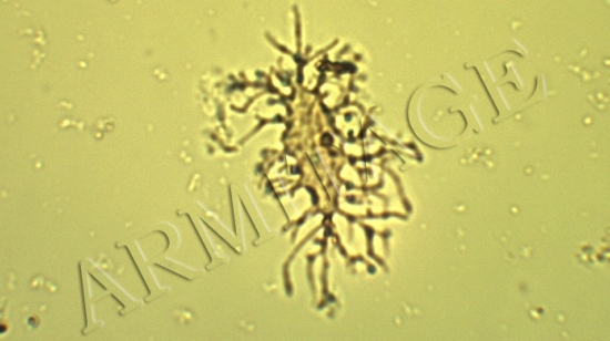
While all of these images are incredible, he saves the best for last. Using a six-week process involving a weak acid, dialysis tubing, and distilled water, he was able to isolate individual bone cells. Look at the photo at the top of this post. It is of an individual Triceratops bone cell, as seen with a standard light microscope. The final two images of Armitage’s paper show two bone cells like the one above. They are free of any surrounding tissue, and one of them shows what appears to be the cell’s nucleus! If I hadn’t been told that these cells came from a Triceratops fossil, I would think they had come from a living animal’s bone tissue.
While the images themselves are stunning, and his accomplishment in isolating individual dinosaur bone cells is quite noteworthy, I found one of his statements to be the most important part of the article:
The remarkable preservation of delicate ultrastructures such as filopodia and cell-to-cell junctions (white arrows, Figures 6 and 7) has resisted a simple explanation despite hypothesized temporal limits on molecular preservation over millions of years. In the case of soft vessels recovered from dinosaur femur specimens, it seems reasonable that these tissues were sequestered from the elements and from biological scavenging activity because of deep encapsulation within compact bone. Within the Triceratops horn, however, which was highly vascular, no sequestration was likely because all of the vessels were openly exposed to air, soil, water, scavengers, dissolved salts and minerals, and the freeze-thaw cycle and heat of Montana seasonal weather; yet a high degree of preservation persists. While plant roots, fungal hyphae, and insect remains were all found traversing the horn, soft fibrillar sheets of bone and well-preserved osteocytes remain.
In other words, for some soft tissue discoveries, such as the work done by Schweitzer and others, you can imagine that the tissues were at least protected from decomposition because they were found inside an intact bone. That wasn’t the case for his specimen, however. Plant roots, along with the remains of insects and fungi, show that the interior of this fossil was exposed to the elements. Nevertheless, soft bone tissue and soft cells from the Triceratops are still there!
Mark Armitage’s work is some of the most important research being done from a creationist perspective right now, and his facility is a unique creationist resource. I hope he can find enough financial support to keep it going!
Good to read. I like Mark. I’ve had a few encounters with him in the web-o-sphere and he seems like a great guy. I can see why he’s so reviled by those seeking to quell Y.E. science.. He’s a very brash, loud, outspoken human with very little padding to his words… but I think that’s what I like.
I’m very pleased to see his hard work and perseverance rewarded and pray that his research continues to be funded by grass roots Christians.
One step closer to dino D.N.A. ? Glad people are still looking.
If nothing else, we should get some good dinosaur proteins out of this research.
So where are the experts from InGen, then? I’m still waiting for the Park to open! Seriously, though, this is truly fascinating stuff!
Here’s a link to download a PDF of Armitage’s article: https://www.researchgate.net/profile/Mark_Armitage2/publication/289686201_Preservation_of_Triceratops_horridus_Tissue_Cells_from_the_Hell_Creek_Formation_MT/links/57ad0e1a08ae3765c3bb0cf7.pdf
I also see from Google Scholar that two other researchers have already cited his paper. Hopefully this will be a spur to secular scientists to go looking for more cells.