Astronomers are Finally Starting to Question an Absurd Assumption

The black circles in this figure represent the 73 quazars that make up the largest structure in the observed universe. The red crosses represent the 34 quazars that make up another massive structure. (Image is from reference 3.)

One of the more absurd assumptions that is routinely made in astronomy is called the cosmological principle. One way to phrase the principle is:

Viewed on a large enough scale, the properties of the universe are the same no matter where you are.

However, observations have never supported this assumption. Instead, the observable universe seems incredibly “lumpy,” with huge structures separated by vast areas devoid of structures. Nevertheless, cosmologists have doggedly taken the cosmological principle as their starting assumption when it comes to developing models of the universe, despite the fact that observations don’t support it.

Indeed, the cosmological principle is a necessary starting point for the Big Bang, which most, but certainly not all, astronomers think is a good description of the origin and development of the universe. As Paul Fleisher says in his book, The Big Bang:1

The cosmological principle is the central idea of the Big Bang theory. This rule says the universe is homogeneous and isotropic at very large scales.

Even if we go away from the Big Bang model, the vast majority of models that attempt to describe the universe start with the assumption that the cosmological principle is valid. There are some models that do not start with that assumption, but they are few and far between.2

I have always been skeptical of the cosmological principle, simply because it isn’t supported by observation. The universe doesn’t look homogeneous at all. Instead, it looks really “lumpy.” Nevertheless, when I read the scientific literature, the cosmological principle seems to be considered a fact in almost all of the astronomy-related papers.

It looks like that might be starting to change.

Continue reading “Astronomers are Finally Starting to Question an Absurd Assumption”

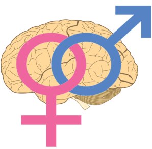
Male DNA in Female Brains? Yes!

Male DNA was found in 63% of the women studied.
(Public domain image)
A gene called DYS14 is found only on the Y chromosome in human beings. Of course, only males have a Y chromosome, so it is reasonable to assume that the only place you will ever find this gene is in men, right? Wrong! William F. N. Chan and his colleagues examined the brains of 59 deceased women, and they found the gene residing in 37 of the brains studied! In other words, 63% of the deceased women studied had male DNA in their brains. Interestingly enough, in most of those brains, the DNA was found in several different places!1

How in the world did male DNA get into these women’s brains? The researchers aren’t sure, because they don’t have detailed medical histories for most of the women. However, the most likely explanation is that the DNA comes from the male children that these women carried. I have written about this phenomenon, called fetomaternal microchimerism, before. As I mentioned in that article, when I first heard about a baby leaving cellular remains inside his or her mother, I thought it couldn’t possibly be true. However, I was wrong. There is solid evidence to suggest that not only do babies leave a lasting, cellular imprint on their mothers, mothers do the same for their babies.

However, the possibility that children leave some of their DNA behind in their mother’s brain is very surprising. After all, the cells that make up the brain are incredibly sensitive. In fact, the contents of your own blood are toxic to your brain cells. As a result, you have an elaborately designed blood-brain barrier that shields your brain cells from your blood. This barrier is so vigilant that it allows only certain substances (such as the glucose and electrolytes that the brain cells need) to pass through it. As a result, your brain cells are protected from the majority of substances found in your bloodstream.2

Continue reading “Male DNA in Female Brains? Yes!”

Bacteria in Breast Milk? Yes!

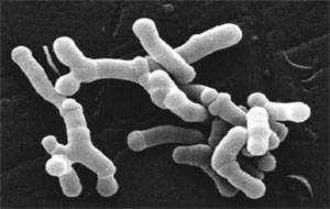
These Bifidobacterium longum bacteria are often found in the intestines of infants. (Click for credit)
The breast milk that a mother feeds her baby is laden with bacteria. Does that sound bad? It shouldn’t! While there are some pathogenic bacteria, most bacteria are incredibly beneficial to the life that exists on this planet. That’s especially true of bacteria that live in and on people. It turns out that most people live in a relationship with more than 150 different species of bacteria, and the individual bacteria that participate in this relationship far outnumber the human cells that make up a person’s body. In one sense, then, a person is not an individual. Instead, he or she is a walking ecosystem!

Scientists now call the collection of bacteria that lives in a person’s body the microbiome, and as the article linked above indicates, each person seems to have his or her own special mix of bacteria in that microbiome. Indeed, some researchers think that analyzing the DNA of the bacteria a criminal leaves behind can aid in identifying that criminal in cases where his or her own DNA is not available at the crime scene or too degraded to analyze properly.1

So where does an infant start collecting the bacteria that will make up his or her own microbiome? One of the sources is the breast milk that the infant drinks. It has been known for quite some time that breast milk contains bacteria, but the details have not been well studied. However, a group of Spanish researchers have begun to shed some light on those details. They studied the breast milk of 18 mothers who varied in weight, weight gain during pregnancy, and the mode in which the baby was delivered. They sampled the milk these mothers produced at three different times: the first secretions of milk produced after giving birth (called colostrum), the milk that was produced one month after giving birth, and the milk that was produced six months after giving birth. They sequenced the DNA of the bacteria found in these samples of milk, and they came up with some amazing results.2

Continue reading “Bacteria in Breast Milk? Yes!”

There is No Such Thing…

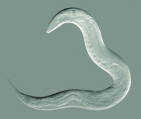
This tiny, transparent roundworm has amazing neurons. (Click for credit)
In my high school textbook, I try to emphasize the fact that there is no such thing as a “simple” life form. Even the most basic living organism is a marvel of amazing complexity. Consider, for example, the tiny roundworm, Caenorhabditis elegans, pictured on the left. It is only 1 millimeter long, and because it is transparent, it is very easy to study. In addition, because it’s nervous system is considered “simple,” it has been examined extensively in order to understand how animal nervous systems work.

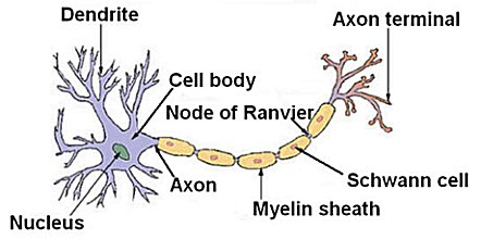
Why is its nervous system considered “simple?” Well, the functional unit of an animal’s nervous system is the neuron, a sketch of which is given below:

A simplified sketch of a neuron (Image in the public domain)

These individual cells receive signals in their dendrites and transmit them through the cell body and down the axon. Most animal nervous systems are made up of many, many neurons. For example, in the part of the brain known as the cerebral cortex, cats have about 300 million neurons, dogs have about 160 million neurons, and chimpanzees have about 6.2 billion neurons. The animal with the largest number of neurons in the cerebral cortex is probably the African elephant, topping off at about 11 billion neurons, but the false killer whale comes in as a close second, at about 10.5 billion. By comparison, the cerebral cortex of a person contains about 11.5 billion neurons.1

The entire nervous system of C. elegans is a mere 302 neurons. That’s really simple compared to people and animals isn’t it? Well…not exactly.

Continue reading “There is No Such Thing…”

What Do Spiders Do With All Those Eyes?

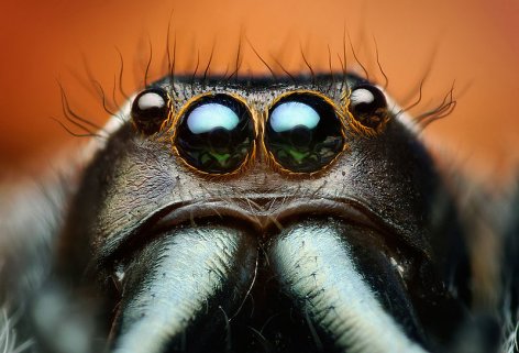
This photo shows four of the eight eyes on a jumping spider. The middle two are the principal eyes, and the other two are the anterior lateral eyes (ALEs). (click for credit)

Most spiders have eight eyes, and their arrangement varies depending on the type of spider. In fact, when studying a spider, scientists often use the number and arrangement of the eyes to help them classify the specimen.1 What does a spider do with all those eyes? Well, in the case of a jumping spider, we know that the two large eyes near the center of the head are the spider’s principal eyes. They can see sharp images, are sensitive to color, and can move to track a target.

The eyes that are right next to the principal eyes are called the anterior lateral eyes (ALEs). They cannot move, do not seem sensitive to color, and as far as we can tell, don’t really allow for the spider to see images. Instead, it has always been thought that these eyes help the spider detect motion.2 But what about the principal eyes? Do they detect motion as well? Three researchers decided to determine the answer to that question by conducting a interesting experiment with some spiders and an iPod touch.

They ended up using removable paint to “blind” specific eyes of jumping spiders from the species Phidippus audax. For 16 of the spiders, they used the paint to “blind” only the principal eyes. They then used the paint to “blind” only the ALEs of 14 other spiders. Finally, they used 16 spiders with none of their eyes “blinded” as a control group. One at a time, they put the spiders in an “arena” that had four walls. Three were foam-core walls, and the fourth wall was the screen of the iPod touch. They allowed each spider to acclimate to the arena and waited for its head to face the screen. When that happened, they remotely started an animation of a black circle either looming towards the spider or retreating from the spider. The results they got were quite interesting.

Continue reading “What Do Spiders Do With All Those Eyes?”

It’s Amazing What RNA Can Do!

A sunburn comes from micro-RNAs that are released by damaged cells. (Click for credit)
One of the truly remarkable things about creation is how one substance can be used in nature to do all sorts of different jobs. Take ribonucleic acid, for example. Commonly referred to as RNA, scientists have known for quite some time that it is an integral part of how the cell makes proteins. A particular kind of RNA, called messenger RNA, copies a protein recipe contained in DNA, and it takes that copy to a protein-making factory called a ribosome. Once the recipe is at the ribosome, two other kinds of RNA, transfer RNA and ribosomal RNA, interact with the messenger RNA to build the protein in a step-by-step manner.

Because RNA is such an important part of how the cell builds proteins, some scientists speculated that this was its only job. In 1993, however, Victor Ambros, Rosalind Lee, and Rhonda Feinbaum found another job for RNA. Short strands of RNA, which are now called microRNAs, sometimes regulate how much of a particular protein is made in the cell.1 Since then, other forms of RNA have also been shown to regulate the amount of protein produced in a cell. In addition, scientists have found that some types of RNA perform functions that aren’t even directly related to the production of proteins. For example, some types of RNA serve as “molecular guides,” taking proteins where they need to be in the cell, while other types of RNA serve as a “molecular adhesives,” holding certain proteins to other RNA molecules or to DNA.

Now even though the last two jobs I mentioned are not directly related to protein production, they still involve proteins. So is it safe to say that while RNA performs several functions in the cell, all of them are related to proteins in some way? I might have answered, “Yes” to such a question if a student had asked me that just a few weeks ago. However, a new paper in Nature Medicine has found a function for some microRNAs that has nothing to do with proteins. Some microRNAs serve as radiation detectors.2

Continue reading “It’s Amazing What RNA Can Do!”

Move Over, Kindle. This Scientist Stored His Book on DNA!

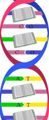
DNA stores information more efficiently than any human technology. (montage of Art from Kevin Spear and the public domain)
Everyone has heard of DNA, but many don’t appreciate its marvelous design. It stores all the information an organism needs to make proteins, regulate how they are made, and control how they are used. It does this by coding biological information in sequences of four nucleotide bases: adenine (A), thymine (T), guanine (G), and cytosine (C). The nucleotide bases link to one another in order to hold DNA’s familiar double-helix structure together. A can only link to T, and C can only link to G. As a result, the two linking nucleotide bases are often called a base pair. DNA’s ingenious design allows it to store information in these base pairs more efficiently than any piece of human technology that has ever been devised.

What you might not realize is that pretty much any information can be stored in DNA. While the information necessary for life involves the production, use, and regulation of proteins, DNA is such a wonderfully-designed storage system that it can efficiently store almost any kind of data. A scientist recently demonstrated this by storing his own book (which contained words, illustrations, and a Java script code) in the form of DNA.1

The way he and his colleagues did this was very clever. They took the digital version of their book, which was 5.27 megabits of 1’s and 0’s, and used it as a template for producing strands of DNA. Every time there was a “1” in the digital version of the book, they added a guanine (G) or a thymine (T) to the DNA strand. Every time the digital version of the book had a “0,” they added an adenine (A) or a cytosine (C). Now unfortunately, human technology cannot come close to matching the incredible design of even the simplest living organism. As a result, while living organisms can produce DNA that is billions of base pairs long, human technology cannot. It can produce only short strands of DNA.2 So while a single-celled organism could have produced one strand of DNA that contained the entire book (and then some), the scientists had to use 54,898 small strands of DNA to store the entire book.

Continue reading “Move Over, Kindle. This Scientist Stored His Book on DNA!”

Animal Magnetism

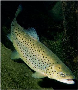
Brown trout like this one return to the stream in which they hatched in order to spawn. (Click for credit)
Many species of fish, such as the brown trout pictured on the left, hatch in streams and then travel away from those streams in order to mature. However, when it is time to reproduce, they end up navigating back to the same stream in which they hatched so they can spawn there. How do they accomplish this? How do they know where they are and which way to swim in order to get back to that special stream? Based on behavioral studies, scientists have thought that these fish are able to sense the earth’s magnetic field and use it as an aid in their navigation. However, the specific source of this magnetic field sense has been elusive…until now.

A recent study has shed a lot of light on this magnetic sense, at least for trout (and presumably other similar fish, like salmon). The authors of the study set out to determine what gives the trout their magnetic sense, and they developed a rather ingenious method to aid them in their search. First, they took tissue samples from the trout’s nasal passages, because previous studies indicated that there was magnetite (a mineral that reacts strongly to magnetic fields) in those tissues.1 Then, they put cells from the tissues under a microscope and exposed the cells to a rotating magnetic field. In response, some of the cells rotated with the field.2 You can actually see a video of this happening here! Just click on the links for downloading the movies.

This is a very simple, very sensitive method for finding the cells responsible for the trout’s magnetic sense. As you can see from the video, the cells that are sensitive to the rotating magnetic field are smaller than the other cells in the tissue. Also, the authors found that only 1 in 10,000 cells in the nasal tissue have a magnetic sense. No wonder these cells haven’t been found until now! Of course, as the authors studied the cells more closely, they found evidence of thoughtful design.

Continue reading “Animal Magnetism”

One Gene = One Protein? Not Even Close!

In a previous post, I discussed alternative splicing, an amazing aspect of our DNA that allows it to store information in a compact, elegant way. In brief, a gene is actually a recipe that the cell uses to make a particular protein. Since most of a cell’s DNA is in the nucleus, the “recipe” stored in that gene must leave the cell’s nucleus in order to be turned into a protein. To do that, the “recipe” is copied by a molecule called messenger RNA (mRNA). The mRNA then takes the copied “recipe” out of the nucleus to the ribosome, which is where proteins are made.

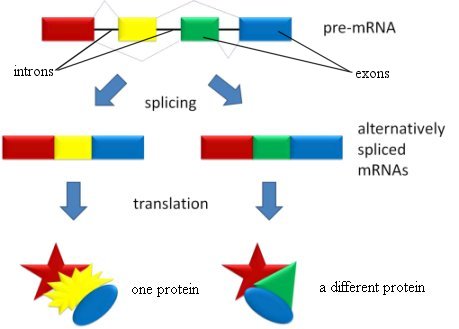
In eukaryotic cells (the kinds of cells found in plants and animals), however, something very interesting happens before the mRNA leaves the nucleus. Some parts of the mRNA are cut away, and the remaining parts are then stitched back together. The parts of the mRNA that are cut away never leave the nucleus, so they are called introns (they stay IN the nucleus). The remaining parts that are stitched together are called exons (they EXit the nucleus). For a while, geneticists didn’t know the purpose of introns, so in typical evolutionary fashion, many decided that they had no purpose, and introns were lumped into the category of “junk DNA.” Of course, as we have learned more about genetics, we have learned that the evolution-inspired idea of “junk DNA” is, itself, junk, although some evolutionists still cling to it.

Nowadays, of course, we know the reason that introns exist. It is part of the design of the Creator, allowing DNA to store information in an incredibly efficient way. Each exon represents a “module” of useful information. If the cell stitches the exons together in one way, it makes one protein. If it stitches the exons together in another way, it makes a different protein. As a result, a single gene can actually produce many different proteins. The introns not only serve as a means by which the cell can identify the exons, they also regulate the amount of the various proteins that are being made.

This process of alternative splicing is illustrated in the figure below:

Because of alternative splicing, a single gene can tell the cell to produce different proteins. (public domain image)

In the previous post about alternative splicing, I discussed how recent evidence suggests that up to 95% of the genes in the human genome participate in this process. However, I did not address how many different proteins a single gene can produce using alternative splicing. In some cases, the answer is truly astounding.

Continue reading “One Gene = One Protein? Not Even Close!”

Another Example of Three-Way Mutualism. Is This Just the Tip of the Iceberg?

A white-spotted pufferfish in a seagrass bed (click for credit)

Over two years ago, I wrote about an interesting three-way mutualistic relationship between a virus, a fungus, and a plant. Less than a year later, I wrote about how people are actually walking ecosystems, participating in a huge number of mutualistic relationships with many different species of bacteria. Last night, while reading the scientific literature, I ran across another example of a three-way mutualistic relationship, and it is equally as fascinating!

This three-way relationship starts with seagrasses. Coral reefs are the “stars” of the marine world, but seagrass communities can be considered its “workhorses.” While they make up only 0.2% of the ocean’s ecosystems, they produce more biomass than the entire Amazonian rainforest!1 Why are they so productive? Because they form a wide variety of marine ecosystems that serve as nurseries for many developing fishes and homes to a wide variety of sea creatures including turtles, manatees, shrimp, clams, sea stars, etc. Because of their amazing ability to support such ecosystems, seagrasses have been studied by marine biologists for some time. However, there has always been a nagging mystery associated with them.

The roots of seagrasses trap sediments which form a rich mud that is often several feet deep. The mud is rich because it contains all manner of decaying organic matter. However, the reason the organic matter decays is because bacteria decompose it. One of the byproducts of this bacterial decomposition is sulfide, and if that sulfide were allowed to build up to high concentrations, it would actually end up harming the seagrasses themselves. However, it never does. No one has proposed a satisfactory explanation as to why this doesn’t happen.

Certainly, the seagrasses transport oxygen to the mud through their roots, and that oxygen can turn the sulfide into sulfate, which is harmless to the seagrasses. However, detailed studies show that the sulfide produced by the resident bacteria accumulates far faster than it can be removed by the oxygen that is added to the mud through the seagrasses’ roots, especially during warm seasons.2 Thus, there must be some other way that sulfide is being removed from the mud.

Marine biologists had no idea what this other way was…until now.

Continue reading “Another Example of Three-Way Mutualism. Is This Just the Tip of the Iceberg?”