
I have a small plaque on my wall that reads, “Docendo Discimus.” It is a Latin proverb that means, “By teaching, we learn.” The very first time I helped a friend with her homework, I realized the truth of that proverb, and it is one of the reasons I enjoy teaching so much. I love to learn, and since teaching helps me to learn, I love to teach. Part of the proverb’s truth comes from the fact that as we explain things to others, we think about them in new ways, which often leads to new insights. Over the years, however, I have found other ways that teaching helps us to learn, and I experienced one of them this past weekend.
Because of the Lord’s leading, I decided to offer some online high-school courses this year. It has been a blast teaching students from all over the U.S. as well as a handful of foreign countries. Along with the joy of teaching and getting to know students, however, comes the drudgery of grading. While I love the former, I most certainly do not love the latter. However, on Saturday, I graded lab reports from my various classes.
I started with my high school biology class, which had spent the last week or so culturing pond water and looking at samples of it under the microscope. A lab like that is “hit or miss,” because finding interesting microscopic creatures in pond water depends on a lot of factors. Some students see many organisms, while other students see few or none at all. I was in the “monotony zone,” having gone through several lab reports, when I came across a submission with an excited note attached. The student, Brynna Taylor, was thrilled with what she saw, and she just had to share a few videos with me.
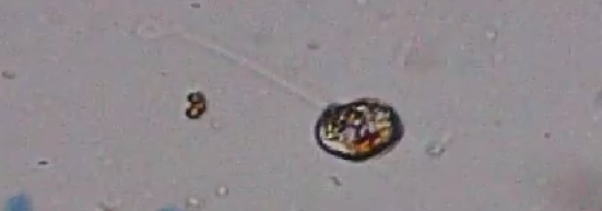
The first of three videos is posted (with her permission) below. She thought the organism she was focusing on was a Euglena, and while I understood her reasoning, I quickly realized she wasn’t correct. Euglena typically have only one flagellum (a tail-like appendage used to move around), and the organism she was focusing on had two. In addition, only one seemed to be used for movement. The other was pushed out in front of the organism, almost like it was using the flagellum to sense what was ahead.
I had never seen anything like that before, so I did some research and came up empty. Then I did something I rarely do. I asked my sister-in-law (who is a biology professor) for help. She has forgotten more biology than I will ever know, and I always learn more about the subject when I visit her and her husband, who is a world-renowned expert in molecular biology. However, they are both very busy people, so I don’t like to bother them unless I have no other options. I sent her the video, and she also couldn’t identify the organism. However, she gave me some ideas on how to refine my search, and I eventually learned that this interesting organism is from genus Anisonema in kingdom Protista, subkingdom Protozoa.
I have probably searched hundreds of drops of pond water under the microscope throughout my life, and I have seen a lot of “cavorting beasties,” as Anton van Leeuwenhoek referred to them when he first saw them in 1673. I thought I had pretty much seen it all, but I didn’t even know about this genus. However, because a student was excited about what she saw, I learned about a genus that was entirely new to me. My sister-in-law did as well. When I asked her to confirm my identification, she said:
That does look like the video. Good work! Protists are so hard — so darn many of them!!
While learning about a genus that is entirely new to me is interesting, I learned something even more valuable from Miss Taylor: God’s creation is so marvelously complex and diverse that there is no way you could see everything there is to see, even when you are just looking at a drop of pond water!
If you would like to see the other videos Brynna Taylor shared with me, they are given below. This one is (most likely) a Euglena:
This last one really demonstrates why Anton van Leeuwenhoek referred to microscopic organisms as “cavorting beasties.” Unlike the previous two videos (which came from a freshwater pond), this comes from a sample of ocean water. Miss Taylor had put an egg in the ocean water to give the microorganisms something to eat, and a slime developed. She put a sample of slime under the microscope, and the video below is the result. There are a lot of different microorganisms there, but the most common ones seem to be paramecia.
I hope you will be teaching those classes again next year–I’d love my ninth grader to be able to take biology with you!
I am listening to the Lord to see. I will announce it here when I decide, which will probably be in February or March.
That’s amazing! Do you know which microscope with a video option she is using? My daughter is in the Tuesday class and I’d love to have a more powerful option for her. Thanks!
She used the Celestron PentaView LCD Digital Microscope. It’s about $150 more than the basic microscope you need for the course, but it obviously has some real benefits!
https://www.amazon.com/Celestron-PentaView-LCD-Digital-Microscope/dp/B007DJZEFC/ref=sr_1_1?ie=UTF8&qid=1508416263&sr=8-1&keywords=Celestron+PentaView+5+MP
My compliments to Brynna for her videos. The image quality is very good on all of them.
I used an older Zeiss scope for many years and decided to pick up one of the cheap Amazon scopes for use in a separate location in my office. I figured it would probably not be very good, but that I could revert back to the Zeiss if I needed better image quality. I bought an AmScope branded scope and was surprised to find it gave me better viewing images than the old Zeiss.
I am retired now so I don’t need a microscope. If I was buying one today, I would consider one of the trinocular scopes that are available because of the ability to use a camera on a separate viewing tube. I’ll paste a link to one that looked interesting to me.
https://www.amazon.com/AmScope-T490B-Magnification-High-Resolution-Microscopes/dp/B004QEFO1Q/ref=pd_sbs_328_4?_encoding=UTF8&psc=1&refRID=8G0F5XQ9EG5PZRPTKC66
Thanks, Bill. That one is actually less expensive than the one the student used, so that’s great.
Yes, it is less expensive, but I don’t think this model comes with a camera. That would be an additional expense. There are other binocular or trinocular scopes out there with built in cameras. The advantage of the one I posted is that you could start with something as simple as a cell phone adapter and then get a better dedicated camera if you find the cell phone is not adequate for your photo or video needs. I’ll paste a link to an example of a cell phone adapter.
https://www.amazon.com/Microscope-Adapter-Smartphone-Camera-Adaptor/dp/B07412S738/ref=sr_1_3?s=electronics&ie=UTF8&qid=1508501385&sr=1-3&keywords=microscope+camera
I would like to add one additional comment for students, or others, who already have scopes without a camera built in to the scope. I participated in two different continuing education cytology classes within the past couple years. The majority of the images posted in those courses, by both instructors and class participants, were taken by simply holding a cell phone up to the eyepiece of the microscope with no adapter. It is a little tricky to learn at first, but there were many good images taken using that simple technique.
The instructors had access to microscopes with built in cameras but liked the convenience of using their personal cell phone cameras for many of their images. In my opinion, an adapter would help with the positioning and stability but good images can certainly be obtained without using an adapter.
Thanks, Bill!