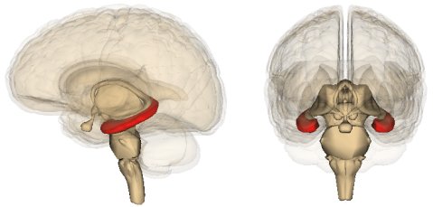
One of the features of the mammalian brain is a structure called the hippocampus. Since the brain is split in half, every mammal has two hippocampi, one on each side, as illustrated in the drawings of the human brain above. These structures are very important for the formation of memories as well as spatial navigation. The reason I am telling you all this is because an incredibly interesting study was just published in the journal Cell, and it uses the aftereffects of the atomic bombs (both their use and testing) to pin down the specifics of how many new brain cells people make in their hippocampi throughout their adult life.
At one time, it was considered a rather strong scientific fact that adult mammals do not produce new neurons (the cells that make up the basic building blocks of the nervous system). For example, An Introduction to Neural Networks (a textbook published in 1995) puts it this way:1
In mammals, although not in many other vertebrates, central nervous system neurons have an important peculiarity; they do not divide after a time roughly coinciding with birth. When a neuron dies, it is not replaced.
Prentice-Hall’s textbook, Exploring Life Science (published in 1997), tells us what this means for people:2
All the neurons you will ever have were formed by the time you were six months old.
We now know that such statements are incorrect. In a variety of mammals that have been studied, adults produce new neurons in the olfactory bulb (a part of the brain used in the sense of smell) and the hippocampus.3 This new study uses a technique that shows adult humans produce a significant number of new neurons in their hippocampi, but they probably don’t produce new neurons in their olfactory bulbs.
When it comes to studying how adult animals produce new neurons (a process now known as neurogenesis), scientists are able to do all sorts of invasive procedures that can’t ethically be done on human beings. As a result, determining the details about human neurogenesis has been much more difficult. However, a team of scientists decided to do something very creative: they used atomic bombs as an indicator of cell age!
How did they do that? Well, from 1945 to 1963, the use and testing of atomic bombs significantly increased the amount of carbon-14 (a radioactive isotope of carbon) in the atmosphere. However, after the Limited Test Ban Treaty of 1963, most above-ground tests of atomic bombs ceased. As a result, the amount of carbon-14 in the atmosphere has been decreasing. Now please note that this decrease is not because of the radioactive decay of carbon-14. That process is too slow to produce a significant change in the past 50-60 years. The decrease has occurred because carbon-14 is being taken out of the atmosphere by living organisms.
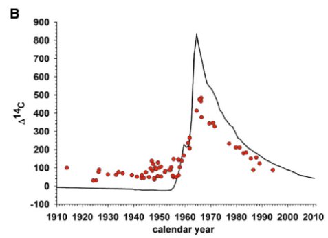
There are two keys for this study. First, when a new cell forms, the parent cell must use carbon from the body to construct a copy of its DNA. Well, that carbon comes from the food we eat, and that food has the same level of carbon-14 as the surrounding atmosphere. Second, the amount of carbon-14 in the atmosphere has been measured for many, many years. As a result, the authors realized that they could correlate the amount of carbon-14 in a cell’s DNA to when it was first produced. For example, here is a graph they show for the carbon-14 content in the DNA of human cells that we know constantly reproduce over time:4

Notice how well the carbon-14 content of the cell’s DNA (the red dots) track the carbon-14 content of the atmosphere (the black line). Because of this nice correlation, the authors were able to use the carbon-14 content in any cell’s DNA to determine the year in which it was produced!
In the study, the authors took brain cells from people who had died. Using this technique, they showed (in a previous study5) that the cells in the olfactory bulbs were produced roughly when the person was born. As a result, they concluded that unlike most mammals, people do not produce new neurons in their olfactory bulbs. However, in the current study (reference 4), they showed that many of the neurons in the hippocampi were much younger than the individuals from whom the cells were taken. As a result, they conclude that people do produce new neurons in their hippocampi throughout their lives.
Now as I have mentioned previously, the fact that adults make new neurons isn’t news. It has been firmly established for a few years now.6 The real news is what this study says about how many are produced. It indicates that adults produce about 700 new neurons in each hippocamus every day!. That’s a lot more than I expected! In addition, they show that the number of new neurons produced per day declines with age. For most of a person’s life, neurogenesis is faster in the hippocampus than it is in the same region of a mouse’s brain, which is probably the best-studied mammalian brain when it comes to this process. In fact, a person has to reach middle age before his or her rate of neurogenesis slows down to what is seen in mice. As the authors state:
We conclude that neurons are generated throughout adulthood and that the rates are comparable in middle-aged humans and mice, suggesting that adult hippocampal neurogenesis may contribute to human brain function.
So this technique shows that there is little adult neurogenesis in the human olfactory bulb (unlike we see in most mammals), but a lot of adult neurogenesis in the human hippocampus. Most likely, these new neurons are important to the function of the hippocampus, and the fact that neurogenesis is so rapid in people compared to other mammals (like mice) is probably related to the fact that people have higher cognitive abilities. In addition, the sense of smell is not as important to people as it is to many other mammals, and that’s probably why other mammals produce new neurons in their olfactory bulbs but people do not.
It will be interesting to see what further studies tell us about adult neurogenesis!
REFERENCES
1. James A. Anderson, An Introduction to Neural Networks, Massachusetts Institute of Technology 1995, p. 6
Return to Text
2. Anthea Maton, Exploring Life Science, Prentice-Hall 1997, p. 656
Return to Text
3. Brus M, Meurisse M, Gheusi G, Keller M, Lledo PM, Lévy F., “Dynamics of olfactory and hippocampal neurogenesis in adult sheep,” Journal of Comparitive Neurology 521(1):169-88, 2013
Return to Text
4. Kirsty L. Spalding, Olaf Bergmann, Kanar Alkass, Samuel Bernard, Mehran Salehpour, Hagen B. Huttner, Emil Bostrom, Isabelle Westerlund, Celine Vial, Bruce A. Buchholz, Goran Possnert, Deborah C. Mash, Henrik Druid, and Jonas Frisen, “Dynamics of Hippocampal Neurogenesis in Adult Humans,” Cell 153:1219-1227, 2013
Return to Text
5. Bergmann, O., Liebl, J., Bernard, S., Alkass, K., Yeung, M.S., Steier, P., Kutschera, W., Johnson, L., Lande´ n, M., Druid, H., “The age of olfactory bulb neurons in humans,” Neuron 74:634–639 2012
Return to Text
6. Amanda Sierra, Juan M. Encinas, and Mirjana Maletic-Savatic, “Adult Human Neurogenesis: From Microscopy to Magnetic Resonance Imaging,” Frontiers in Neuroscience 5:47, doi:10.3389/fnins.2011.00047, 2011
Return to Text
