Counting To God: Another Atheist Who Became a Christian

Douglas Ell, MIT graduate and former atheist (click for credit)
Douglas Ell, MIT graduate and former atheist
(click for credit)
Douglas Ell graduated from the Massachusetts Institute of Technology (MIT) with degrees in math and chemistry. He then went to the University of Maryland, where he earned a Master’s degree in theoretical mathematics. Not satisfied with only three degrees, he also went to law school and graduated magna cum laude. After that, he began his career as an attorney.

When he was a child, he went to church, but the older he got, the less he believed in God. By the time he was in high school, he wrote to his minister and stated that he no longer believed in God. His minister wrote back and gave him a book to read, but Ell never read it. By the time he got his law degree, he was a full-fledged atheist. In his new book, Counting To God, he describes what he believed at that point in his life:

It seemed you could explain just about everything with logic and science. It seemed God had no place in our modern world. I treated God like a joke. (p. 19)

In his early thirties, Ell had a son, and this caused him and his wife to start attending church. Ell treated it like a social club, but he did notice something: Many of the people in the church he attended (including the minister) had an inner peace that he could sense. He wanted that peace, but didn’t see how he could have it, because he didn’t believe what they believed.

In his mid-forties, a new career opportunity forced him to spend a lot of time on airplanes. As a result, he started reading about science, mathematics, and religion. The more he read, the more he saw a connection between the three. He eventually saw seven specific ways in which science and mathematics support the existence of God:

1. The evidence that the universe had a beginning
2. The apparent “fine tuning” of the universe
3. The complexity of life and our inability to discover a naturalistic explanation for its origin
4. The fantastic, futuristic technology that exists in all of life
5. The mounting evidence against neo-Darwinian evolution
6. The specialness of earth
7. The mathematical nature of the universe

Continue reading “Counting To God: Another Atheist Who Became a Christian”

Research Shows That We’ve Been Wrong About Stem Cells

This is a simple schematic of a tooth. (click for credit)
Stem cells are a hot topic in biology. Scientists call them “undifferentiated,” because they have not yet specialized to become a specific kind of cell. This means that a stem cell can develop into several different kinds of cells, depending on what the body needs. For example, everyone has stem cells in their bone marrow. Some of those cells (called hemopoietic stem cells) can develop into various kinds of blood cells, while others (called stromal stem cells) can develop into fat cells, bone cells, or cartilage cells. Physicians have used such stem cells to treat certain heart conditions1, and it is expected that as time goes on, more stem-cell-based treatments will be developed.

Of course, bone marrow isn’t the only place in which stem cells reside. In fact, stromal stem cells can also be found in tooth pulp, the soft tissue that is under the tooth’s dentin (see the illustration above). That’s where the blood vessels and nerves of the tooth are found. While scientists have known for a long time that these stem cells are there, how they get there has always been a mystery.

Nina Kaukua and her colleagues weren’t trying to solve that mystery. They were just studying certain kinds of cells in the teeth of mice. These cells, called “glial cells,” are support cells that help the nerve cells (called neurons) do their job. In their research, they were adding a fluorescent chemical to these cells and watching what happened to them over time. What they found was kind of shocking!

Continue reading “Research Shows That We’ve Been Wrong About Stem Cells”

More Than A Marksman

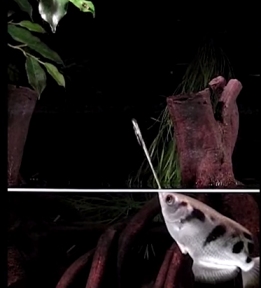
An archerfish hunting a cricket (click for video)
I have been intrigued by archerfish (genus Toxotes) ever since I saw them at an aquarium. They like to feed on insects that crawl around on the plants near the water’s edge. When an archerfish spies an appetizing insect, the fish shoots a stream of water out of its mouth, hitting the insect and knocking it into the water. The fish then goes to the surface and swallows the insect. You can watch a video of this happening by clicking on the picture. Youtube has several other videos of these incredible fish.

Obviously, the archerfish has to “know” a lot of physics to be able to hunt the way it does. After all, as soon as the water leaves its mouth, it is affected by gravity. As a result, the stream of water doesn’t travel straight to its target. Its path bends downward, forming a shape called a parabola. Because of this, the archerfish can’t aim directly at its prey. Instead, it has to aim above its prey, taking the curved shape of the water’s path into account.

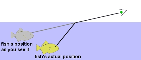
But that’s not the end of the story. When light passes from one medium to another, it bends in a process called refraction. This causes a problem for what we see when we look at things that are in the water. Consider, for example, looking at a fish that is swimming in a pond. You see the fish because light hits the fish, reflects off the fish, and travels to your eyes. However, when the light passes from water into air, it bends, and that causes a problem for you. Look at the drawing below:

refraction

The light coming from the fish bends when it enters the air, but your brain interprets light as traveling in a straight line. So when your brain constructs the image of the fish, it doesn’t take refraction into account, and therefore it forms the image of the fish at a shallower depth and behind where the fish actually is. Those who try to spear fish while standing in shallow water have to account for this. If they don’t aim their spear in front of the place where they see the fish, they will never hit it.

The archerfish, of course, has a similar problem. The light that its eyes receive bends when it hits the water. Because of the way it bends, the fish sees the insect closer and lower than it really is. So not only does the archerfish have to account for the effects of gravity when it aims its water stream, it also has to realize that it shouldn’t aim for the position where it sees the insect. Instead, it should aim for a position that is closer and lower!

If all that isn’t impressive enough, scientists have recently found out that the archerfish uses even more physics when it hunts!

Continue reading “More Than A Marksman”

Confirmation of a Creationist Prediction Becomes Even More Stunning

A model of the vertebrate retina, showing the Müller cells (image by Dr. Jens Grosche, Universität Leipzig, found in reference 2)

Nature is filled with amazing designs, which leads me to the conclusion that it is the product of a Magnificent Designer. Of course, many scientists disagree with that conclusion, and some of them try to argue against it by pointing out examples of what they think are bad designs in nature. One of the oft-cited examples is the retina of the human eye. As Dr. Michael Shermer puts it:1

The anatomy of the human eye shows that it is anything but “intelligently designed.” It is built upside down and backwards, with photons of light having to travel through the cornea, lens, aqueous fluid, blood vessels, ganglion cells, amacrine cells, horizontal cells, and biploar cells, before reaching the light-sensitive rods and cones that will transform the signal into neural impulses.

To understand what he is saying, look at the illustration above. When light hits the surface of the eye’s retina, it has to travel through layers of cells that essentially connect the retina to the rest of the nervous system. Only then can it reach the light-sensitive cells, called rods and cones, and be converted into a signal that can be sent to the brain. This, of course, seems backwards to most evolutionists. According to them, if the retina were designed intelligently, the rods and cones would be at the retinal surface so they are the first thing the light hits. That way, the connecting neurons could be placed behind the rods and cones so they don’t interfere with the light in any way.

Like most arguments inspired by evolution, the more we learned about the human retina, the less reasonable this argument became. Back in 2007, a study published in the Proceedings of the National Academy of Sciences of the USA showed that light doesn’t have to travel through the connecting neurons to reach the rods and cones. Instead, as shown in the illustration above (which appeared on the cover of the journal), there are special cells, called Müller cells, that collect the light and guide it to the rods and cones.2

Three years later (in 2010), an analysis published in Physical Review Letters concluded:3

The retina is revealed as an optimal structure designed for improving the sharpness of images.

The authors of the analysis showed that the position of the rods and cones in the retina combined with the way the Müller cells guide the light to them make them much less sensitive to light that is scattered within the eye itself. This, in essence, reduces the “noise” that the rods and cones would get from errant photons, making the overall image sharper and clearer.

I blogged about this previously, pointing out that it is precisely what creationists predicted and quite opposite what evolutionists maintained. I am bringing it up now because further research has confirmed the creationist prediction in an even more stunning way!

Continue reading “Confirmation of a Creationist Prediction Becomes Even More Stunning”

Geneticists’ Bias Causes a Big Mistake

This is one way to visualize a coding section of RNA.  It has a start codon that tells the cell to start making a protein, followed by a recipe for that protein.  Then there is a stop codon, to tell the cell that it is done making the protein.
This is one way to visualize a coding section of RNA. It has a start codon that tells the cell to start making a protein, followed by a recipe for that protein. Then there is a stop codon, to tell the cell that it is done making the protein.

You’ve heard it many times before. The vast majority of DNA is junk. Of course, the ENCODE project showed how wrong that notion is. Now that we know the vast majority of DNA is functional, you might wonder how in the world the idea of “junk DNA” became so popular among scientists. I suspect there are many reasons, but some recent research has revealed one of them – a bias regarding what it means for DNA to be functional. The research was done on molecules called long non-coding RNAs, which are commonly referred to as lncRNAs.

What are lncRNAs? Well, let’s start with what RNA is. The genes that your body uses are in your DNA, most of which is found in the control center of the cell, called the nucleus. In order for your cells to use those genes, they must be copied by another molecule. This process is called transcription, and the molecule that performs transcription is RNA. Once it has transcribed the gene, RNA leaves the nucleus, at which point it is often referred to as messenger RNA (mRNA), because it is sending a message to the cell.

What’s the message? It is a recipe for building a protein. That recipe is put together in informational units called codons, and it goes to a ribosome, which is a protein-making factory in the cell. The ribosome reads the codons, translating them one-by-one into a protein. Not surprisingly, this process is called translation. How does the ribosome know when to start building the protein? There is a start codon that tells it to start. How does it know when to stop building the protein? There is a stop codon that tells it to stop. As a result, you can think of messenger RNA in terms of the illustration above – it contains a start codon, a recipe for a protein (the blue bar in the illustration), and a stop codon.

So how does this relate to lncRNAs? Well, messenger RNA is referred to as “coding RNA,” because it codes for the production of proteins. LncRNAs are called “non-coding RNAs,” because it was thought that they do not code for proteins. Now there are lots of RNAs that are thought to be non-coding, but lncRNAs are relatively long. That’s how they get their name. Well, it turns out for at least some lncRNAs, every part of their name (except RNA) is wrong.

Continue reading “Geneticists’ Bias Causes a Big Mistake”

The Appalachian Home Educators Conference

The view from Charlie's Bunion, which is a rock formation on the Appalachian Trail.
The view from Charlie’s Bunion, which is a rock formation on the Appalachian Trail.

This past weekend, I spoke at the Appalachian Home Educators Conference. I gave a total of eight talks over three days, which is a lot! Six of the talks were solo: Being a REAL Environmentalist, Why Homeschool Through High School, What About K-6 Science?, “Teaching” High School at Home, “Teaching” the Junior High and High School Sciences at Home, and Teaching Critical Thinking. I also did two talks with Diana Waring: Homeschooling: The Environment for Genius and Textbook Myths and How to Deal with Them.

In addition to having a great time talking with homeschoolers, I got a chance to spend some time in the Great Smoky Mountains National Park. The highlight was an 8-mile hike (4 miles there and 4 miles back) on the Appalachian Trail to a rock formation known as “Charlie’s Bunion.” The rock formation itself isn’t all that spectacular, but the view from it is! The picture above gives you some idea of what I saw. It was truly gorgeous.

Of course, the conference was the reason I went, so let’s get back to that. The talks went well, and I got a lot of great questions. One student who had used some of my books and then went to a secular university came up to me while I was at my publisher’s booth. He had a whole list of questions he wanted to ask me after spending a year learning science from an evolutionary point of view. I enjoyed answering his questions, and I was so happy that he was willing to take the time to get a different opinion instead of just blindly accepting what his professors told him, as is (unfortunately) the case for so many university students.

Continue reading “The Appalachian Home Educators Conference”

Even More on Slime Molds

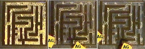
A slime mold chooses the shortest path to the food (labelled "AG") in a maze.  (Image is from the article being discussed.)
A slime mold chooses the shortest path to the food (labelled “AG”) in a maze.
(Image is from the article being discussed.)

I recently ran across a 14-year-old study that I found incredibly interesting. I normally discuss studies that have occurred in the past few years on this blog, but since this study goes well with two other blog posts I have written (here and here), I thought I would go ahead and write about this one as well. In the experiment, the authors studied a slime mold (Physarum polycephalum) that was in its plasmodium stage. During this stage of its life, it is a huge, single cell that has thousands of nuclei.

The researchers grew the slime mold on a maze (as shown on the far left of the image above). Once it had covered the maze, they put out some food in the form of nutrient-rich agar (labelled “AG” in the image above). They put one source of food at the beginning of the maze and another source of food at the end. There were four paths through the maze that connected the two food sources. In a mere four hours, the slime mold had built connections between the two sources using all four of the paths (as shown in the middle of the image above). However, in another four hours, it had worked out the shortest of the four routes (as shown on the right of the image above), and that’s the only one it maintained.

Now as I pointed out in my other two posts about slime molds, these organisms are thought to be “primitive,” because they are thought to have evolved long ago, even before plants and animal evolved. Nevertheless, when presented with puzzles, they are able to solve them. In fact, in one of the previous studies I wrote about, it was suggested that these “primitive” organisms could help us design better networks. Based on the results of this study, such networks would probably be very efficient.

In my high-school biology course, I stress over and over again that there is no such thing as a “simple” organism. The more we study nature, the more clear that becomes.

Yet Another Failed Evolutionary Prediction

This a colony of coral from the genus Acropora, the same genus analyzed in the study that is being discussed.  (click for credit)
This a colony of coral from the genus Acropora, the same genus analyzed in the study that is being discussed. (click for credit)

One of the main ways to test the validity of a scientific hypothesis is to use that hypothesis to make predictions. If those predictions are confirmed by the data, more weight is added to the validity of the hypothesis. If those predictions are falsified by the data, the validity of the hypothesis should be called into question. When it comes to the hypothesis of evolution (in the flagellate-to-philosopher sense), prediction after prediction has been falsified (see here, here, here, here, and here, for example). A recent study published in the Proceedings of the National Academy of Sciences adds to the very long list of failed evolutionary predictions.

In this case, the researchers were studying the phenomenon of apoptosis, which is programmed cell death. In an organism that is composed of several cells, it is important to have a mechanism by which cells that are diseased, very old, or otherwise unstable can be removed. That way, they won’t harm the rest of the organism. This is one of the purposes of apoptosis. When a cell recognizes that it is a potential threat to the organism as a whole, it can actually release protein-destroying chemicals that cause it to kill itself.

Not surprisingly, the process by which apoptosis occurs is incredibly complex. Nevertheless, scientists have made a lot of progress in understanding it. We now know that there are specialized enzymes that start the process. They belong to a group called the TNF receptor-ligand superfamily. In this superfamily, there are TNF ligands (collectively called TNFSF) and receptors (collectively called TNFRSF). When the ligands bind to the receptors, a process starts that can either cause the cell to override its programmed cell death or continue on with it, depending on other chemical signals that are taking place within the organism.

Now don’t get lost in the terminology here. The idea is that multicelled organisms must have a way to get rid of cells that might be bad for the organism as a whole. One way this happens is for special chemicals from a group called TNFSF to bind to other special chemicals from a group called TNFRSF. This activates a process that determines whether the cell should continue to be a part of the organism or kill itself for the good of the organism.

The researchers who published this study decided to analyze apoptosis in one of the more “primitive” organisms on the planet, a species of coral called Acropora digitfera. According to the researchers, corals like this species have been around for 550 million years, so it should be a good representative of some of the earliest animals that ever existed on the planet. Given that assumption, the researchers thought that the apoptosis process in corals should be rather simple – at least a lot less complicated than what we see in the “higher” animals such as flies, birds, and people. Surprisingly, they found the exact opposite.

Continue reading “Yet Another Failed Evolutionary Prediction”

How Do You Design the Best Train? Copy the Designs of the Ultimate Engineer.

This is an Azure Kingfisher.  The Shinkansen "bullet train" in Japan was improved by copying the design of a kingfisher's beak.  (click for credit)
This is an Azure Kingfisher. The Shinkansen “bullet train” in Japan was improved by copying the design of a kingfisher’s beak. (click for credit)

I ran across a story on biomimicry a few days ago. Although it discusses things that happened a while ago, I thought it was a great example of how copying designs found in nature can improve the designs produced by modern science and technology. The story involves Eiji Nakatsu, a Japanese engineer who worked on the high-speed “bullet” trains in Japan. These trains travel at speeds approaching 200 miles per hour, and not surprisingly, there are a lot of design challenges involved in such systems.

In particular, there were three design issues that plagued the trains. First, the train would produce a very loud noise when entering a tunnel, because it would be “smashing” into a column of confined air. While this slowed down the train a bit, the big problem was the noise that it produced. The loud bang would disturb not only wildlife but also nearby residents. In order to comply with Japanese noise pollution regulations, something needed to be done.

According to the article, Nakatsu met this challenge by redesigning the front of the train. As a bird-watcher, he had observed Kingfisher birds diving into water without producing much of a splash. He realized that this was similar to what the trains had to do when entering a tunnel, so he designed the front of the train to be more like the head and beak of a kingfisher. It worked. The train could enter tunnels at full speed without producing a loud noise.

Continue reading “How Do You Design the Best Train? Copy the Designs of the Ultimate Engineer.”

Really Generous Bacteria!

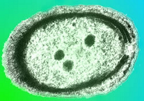
This is an electron microscope image of a bacterium from genus Prochlorococcus.  The colors were added artificially. (click for credit)
This is an electron microscope image of a bacterium from genus Prochlorococcus.
The colors were added artificially. (click for credit)

The image you see above is of a tiny bacterium from genus Prochlorococcus. It is part of a phylum of bacteria called Cyanobacteria, and the members of this phylum are an incredibly important part of the world’s ecosystems. They live in water, converting sunlight and carbon dioxide into sugar and oxygen via photosynthesis. Estimates indicate that cyanobacteria are responsible for producing about 20 to 30 percent of the earth’s oxygen supply.

Prochlorococcus are particularly important cyanobacteria. They are thought to be the most abundant photosynthetic organism on earth, with an estimated worldwide population of an octillion (1,000,000,000,000,000,000,000,000,000).1 More importantly, they tend to live in parts of the ocean that are nutrient-poor. Their photosynthesis helps to alleviate this problem, of course, making them a food source for other organisms that might try to live there.

Dr. Sallie Chisholm at the Massachusetts Institute of Technology (MIT) first described the organisms in 1988 and has continued to study them over the years. She and her colleagues were recently looking at them under an electron microscope and noticed what she described as, “these pimples – we call them ‘blebs’ – on the surface.”2 Dr. Steven J. Biller, a microbiologist who is also at MIT, recognized the blebs as vesicles, which are tiny “sacs” made by nearly every cell in nature. Since the vesicles were found on the surface of the cell, the scientists decided the bacteria were using them to get rid of whatever was inside the vesicles.

They studied the water from their laboratory samples and found that it was, indeed, rich with vesicles that had been released by the Prochlorococcus, and they were surprised by what they found inside.

Continue reading “Really Generous Bacteria!”