What Do Spiders Do With All Those Eyes?

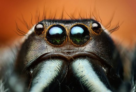
This photo shows four of the eight eyes on a jumping spider. The middle two are the principal eyes, and the other two are the anterior lateral eyes (ALEs). (click for credit)

Most spiders have eight eyes, and their arrangement varies depending on the type of spider. In fact, when studying a spider, scientists often use the number and arrangement of the eyes to help them classify the specimen.1 What does a spider do with all those eyes? Well, in the case of a jumping spider, we know that the two large eyes near the center of the head are the spider’s principal eyes. They can see sharp images, are sensitive to color, and can move to track a target.

The eyes that are right next to the principal eyes are called the anterior lateral eyes (ALEs). They cannot move, do not seem sensitive to color, and as far as we can tell, don’t really allow for the spider to see images. Instead, it has always been thought that these eyes help the spider detect motion.2 But what about the principal eyes? Do they detect motion as well? Three researchers decided to determine the answer to that question by conducting a interesting experiment with some spiders and an iPod touch.

They ended up using removable paint to “blind” specific eyes of jumping spiders from the species Phidippus audax. For 16 of the spiders, they used the paint to “blind” only the principal eyes. They then used the paint to “blind” only the ALEs of 14 other spiders. Finally, they used 16 spiders with none of their eyes “blinded” as a control group. One at a time, they put the spiders in an “arena” that had four walls. Three were foam-core walls, and the fourth wall was the screen of the iPod touch. They allowed each spider to acclimate to the arena and waited for its head to face the screen. When that happened, they remotely started an animation of a black circle either looming towards the spider or retreating from the spider. The results they got were quite interesting.

Continue reading “What Do Spiders Do With All Those Eyes?”

Same Chemical, Different Chemical Formula?

In my previous article, I discussed a chemical found in both the great orange tip butterfly and the marble cone snail. I made the statement that the researchers were surprised to find that the chemical was identical in both species. A commenter asked a good question: When would the same chemical be different (across species or not)? I thought the best way to answer that question was with a new post.

When most of us think about chemicals, we think about simple molecules like water: H2O. The chemical formula of water tells us that there are two hydrogen atoms linked to one oxygen atom. In a glass of water, there are all sorts of molecules like this, and they are all identical. If we do something to change the chemical formula of the molecule, we come up with a completely different chemical. For example, if I were to add one more oxygen to the molecule, I would get H2O2, which is hydrogen peroxide. It is utterly different from water, so in molecules like these, even a change of one atom makes a world of difference.

However, the biological world isn’t quite the same. The molecules are incredibly complex, often composed of thousands of atoms. Consider, for example, proteins. These are large molecules made by linking smaller molecules, called amino acids, together. When amino acids link up together in a specific way, they tend to make a specific protein. An example would be the protein known as cytochrome c. It is a relatively simple protein found in almost all living organisms. It is simple because, as proteins go, it is rather small. In most living organisms, cytochrome c is composed of “only” about a hundred amino acids.1 That might sound like a lot, but there are proteins in living organisms that are composed of more than 25,000 amino acids!2 So as proteins go, cytochrome c is rather “simple.”

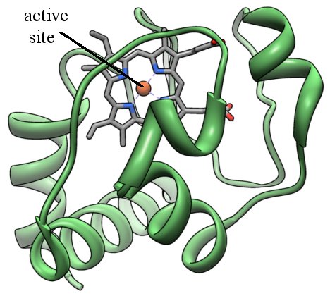
There are many ways to picture a protein, but one way is called a “ribbon diagram.” In this way of picturing a protein, you get a three-dimensional view of the overall backbone of the protein. Here is the ribbon diagram for cytochrome c:

The ribbon diagram of cytochrome c, with the active site pointed out (Click for credit)

The green ribbons represent the structure of the backbone of the protein, and they are composed of many amino acids linked together. The gray bars represent the active site, which is where the protein does most of its work.

Continue reading “Same Chemical, Different Chemical Formula?”

Did Butterflies Evolve from Sea Snails?

The great orange tip butterfly has a toxin in its wings that is identical to the toxin used by the marble cone snail. (Click for credit)

A former student of mine recently alerted me to a study that was published in the Proceedings of the National Academy of Sciences. The authors were studying the proteins found in the wings of a great orange tip butterfly, Hebomoia glaucippe. As they sorted through what they found, they were surprised to find a toxin known as glacontryphan-M.1 The fact that it is a toxin wasn’t surprising to them. After all, Monarch butterflies have cardiac glycosides in their bodies, which are toxic to many birds.2 It is thought that this is a defense mechanism, because birds that eat a monarch butterfly and get sick are unlikely to eat more monarch butterflies.

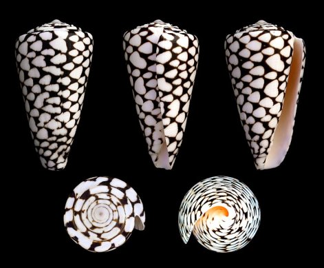
Here’s what’s surprising: the toxin is also found in a sea snail known as the marble cone snail, Conus marmoreus.3 You can see how it gets its name:

The marble cone snail (Click for credit)

The marble cone snail uses the toxin for hunting. It injects the toxin into its prey, paralyzing it. That makes the prey very easy to eat. Obviously, the researchers were surprised to find the same toxin in two separate species that are supposed to be distantly related in terms of evolution. More importantly, they were surprised at the fact that the two toxins are chemically identical.

Continue reading “Did Butterflies Evolve from Sea Snails?”

Desperately Seeking Innovation

One of the biggest problems facing evolutionists is the explanation of how brand new information can be added to a genome. After all, if flagellates eventually evolved into philosophers, an enormous amount of truly original information had to be added to flagellates’ (and their descendents’) genomes. However, genomes are so well-designed and highly-structured, it is difficult to imagine a naturalistic process that could add information to them. Nevertheless, evolutionists have tried their best. One of the more popular notions is gene duplication followed by mutation. We know that genes can be duplicated. It happens quite frequently. The thought is that when a gene is duplicated, one of the copies can continue to produce the protein it is supposed to produce, while the other copy is free to mutate and find some completely new function.

While the thinking behind this idea is logical, experimental evidence to support it has been hard to find. As a result, evolutionists tend to jump on any experimental finding that might suggest the idea is accurate. This is well illustrated by an article that was linked by a commenter on a previous thread. The article claims that researchers have finally shown how a gene can pick up a brand new function, which can then be amplified and modified over time.

Unfortunately, the article’s claim is not accurate. I had already read the scientific paper on which the article was based1, so when I read the article, I understood how incorrect its claims are. However, I am sure the commenter and many other readers of Science Daily do not. As a result, I want to discuss the study’s actual findings. They are very interesting, and they tell us a lot about the genetics of bacterial adaptation. However, they don’t tell us anything about how genes acquire brand new functions or about how information can be added to a genome.

Continue reading “Desperately Seeking Innovation”

Remains of Cells: In DINOSAUR Bones!

New evidence indicates that proteins and DNA still exist in preserved Tyrannosaurus rex bone cells (Click for credit)
In 2005, Dr. Mary Schweitzer stunned the scientific community by publishing data that indicated she had found soft tissue in a Tyrannosaurus rex fossil that is supposed to be more than 65 million years old.1 While many in the scientific community were unconvinced at the time, several lines of evidence now indicate that she was correct. Since that time, other examples of soft tissue in fossils that are supposed to be millions of years old have been found: muscle tissue in a salamander fossil that is supposed to be 18 million years old, retinal tissue in a mosasaur fossil that is supposed to be 70 million years old, and what appear to be bone cells from the same mosasaur fossil. Now, Dr. Schweitzer has come back into the picture with some strong evidence that she has also found bone cells in her Tyrannosaurus rex fossil, as well as one other dinosaur fossil.2

There are three different kinds of bone cells in vertebrates: osteoblasts, osteoclasts, and osteocytes. If you use a microscope, you can tell them apart just by looking at them. Osteoblasts are the cells that build bone, while osteoclasts are the cells that break down bone. Both are important, because your bones adjust to the needs of your body, so there are times that you will need to build more bone, and there are other times you will need to break down some bone. The third group of bone cells, osteocytes, are the most common. They maintain the bone.

The study that found bone cells in a mosasaur fossil found osteocytes, and that’s what Dr. Schweitzer’s team found as well. Now, of course, just because they found microscopic structures that looked like osteocytes isn’t necessarily surprising. After all, the fossilization process could be detailed enough to preserve the shapes of individual cells. If these structures really are just the fossilized shapes of the osteocytes, it is exciting, but not incredibly surprising. However, Schweitzer’s team has done some detailed experiments to show that these aren’t just shapes. Indeed, these osteocyte structures still contain proteins and probably even DNA!

Continue reading “Remains of Cells: In DINOSAUR Bones!”

Remember That Nuclear Disaster in Japan?

Satellite image taken on March 16, 2011, showing reactors at the Fukushima Daiichi Nuclear Power Plant leaking radioactive gas into the air.

On March 11 of 2011, the most powerful earthquake known to have hit Japan struck near the east coast of Honshu. The earthquake generated a tsunami that reached a height of more than 130 feet. Just last month, the Japanese National Police agency reported that there were at least 15,870 people who died, an additional 6,114 who were injured, and 2,814 who are still missing as a result.1 Obviously, it was a disaster of truly stunning proportions.

One of the many things that happened as a consequence of the disaster is that some of the reactors at the Fukushima Daiichi Nuclear Power Plant went into meltdown, and radioactive substances were leaked into the ocean and released into the air. People in a 12-mile radius around the power plant were evacuated so that they would not be exposed to too much radiation. As a result of the meltdown, there is increasing political pressure for Japan to end its reliance on nuclear power. According to the Christian Science Monitor, Prime Minister Yoshihiko’s party has recommended that Japan phase out all nuclear power by the year 2030.

Back when the nuclear disaster was in the news, I commented on it (here and here). Since then, I have been following the scientific literature to see what those who have been monitoring the situation are saying regarding its long-term effects. Recently, a study and some commentary on the study were published in the journal Energy & Environmental Science, and they are surprising, to say the least.

Continue reading “Remember That Nuclear Disaster in Japan?”

Bugs and Bacteria Working Together

A beanbug, Riptortus pedestris (click for credit)
Mutualistic symbiosis, the process by which organisms of different species interact so that all of them benefit, is a very common phenomenon in creation (see here, here, here, here, and here for a few examples). A recent study in the Proceedings of the National Academy of Sciences, USA highlights a very interesting case of mutualistic symbiosis that not only has some important implications for farmers, but also relates to the creation/evolution controversy.

The study examined insecticide resistance in bean bugs (Riptortus pedestris) and similar insects. The authors considered one of the most popular insecticides used by farmers across the world, fenitrothion. It has been known for some time that certain insects, such as the bean bugs in the study, can develop resistance to that insecticide. This is a problem, since bean bugs not only damage bean crops, but also some fruit crops.1 The authors were interested in what causes this insecticide resistance. As they state in the introduction to their paper:

Mechanisms underlying the insecticide resistance may involve alteration of drug target sites, up-regulation of degrading enzymes, and enhancement of drug excretion, which are generally attributable to mutational changes in the pest insect genomes.

In other words, when an insect develops resistance to an insecticide, it is generally assumed that there was a change in the DNA of the insect. A mutation might have damaged the site where the insecticide is supposed to bind; the activity of a gene involved in the destruction of unwanted chemicals might have been enhanced so that the insect destroys the insecticide; or perhaps the activity of a gene involved in getting rid of waste is enhanced so that the insect just excretes the insecticide.

The authors show that for the specific case of fenitrothion resistance in bean bugs and similar insects, none of these mechanisms play a role.

Continue reading “Bugs and Bacteria Working Together”

An Excellent Quote

The cover of Fodor and Piattelli-Palmarini's book
Not long ago, Dr. Jerry Alan Fodor (a professor of Philosophy) and Dr. Massimo Piattelli-Palmarini (a professor of cognitive science) wrote a book entitled What Darwin Got Wrong. I haven’t read the book, but what I have read about it indicates that the authors strongly believe Darwin was right when it comes to the idea that all species descended from a common ancestor. However, they strongly disagree with the mechanism that Darwin proposed (and most Neo-Darwinists accept) for the process by which that happened. While most modern evolutionists contend that mutation acted on by natural selection is the main process by which species adapt and change, the authors argue that it is only one of many considerations. In fact, they go a step further and claim that there is no scientific reason to elevate natural selection above these other processes which it comes to their relative importance.

Since I have not read the book, I cannot comment on the validity of their arguments. However, I ran across a quote from the introduction that makes me want to read the entire book. They call their introduction “Terms of Engagement.” After laying out the terms and outlining the contents of the book, the authors write:

So much for a prospectus. We close these prefatory comments with a brief homily: we’ve been told by more than one of our colleagues that, even if Darwin was substantially wrong to claim that natural selection is the mechanism of evolution, nonetheless we shouldn’t say so. Not, anyhow, in public. To do that is, however inadvertently, to align oneself with the Forces of Darkness, whose goal it is to bring Science into disrepute. Well, we don’t agree. We think the way to discomfort the Forces of Darkness is to follow the arguments wherever they may lead, spreading such light as one can in the course of doing so. What makes the Forces of Darkness dark is that they aren’t willing to do that. What makes science scientific is that it is.

[Jerry Fodor and Massimo Piattelli-Palmarini, What Darwin Got Wrong, Farrar, Straus, and Giroux, First American Edition 2010, p. xx]

I couldn’t agree more. When those who call themselves scientists want to shut off debate on an issue, they are exposing themselves for what they are: rabidly anti-science. Science is all about following the evidence, regardless of where that evidence might lead. It is unfortunate that some (if not many) in the scientific community attempt to silence those who are simply trying to follow the evidence.

Direct Evidence that a Father’s Age Affects Autism Risk

A magnified image of stained human sperm (click for credit)
While there is some disagreement on the subject, most medical scientists would agree that Autism rates are on the rise in the U.S. and in many other parts of the world. What’s the reason for this increase? Like most medical issues, there are probably a variety of reasons. Some have suggested that the increase in autism can be linked to childhood vaccination, but the data argue strongly against it. Most likely, there are a series of genetic and environmental factors that play a role in the increase.

For quite some time now, there has been strong evidence that the age of the father has a significant effect on the chance of his child having autism.1 There has been evidence that the mother’s age also plays a role, but its effect is much smaller.2 However, these studies simply demonstrate a correlation between parental age and autism. They do not show that increased parental age plays a direct role in the cause of autism. However, a recent study published in the journal Nature has changed that. It seems to provide a direct link between the age of the father and autism in the child.

The authors of the study examined the entire genomes of 78 parent-offspring trios (mother, father, and child) to directly determine what mutations the child received from the father’s sperm cell and what mutations the child received from the mother’s egg cell. Because they were specifically interested in the cause of neurological disorders, they used a large number of trios that contained a child with either autism or schizophrenia. In the end, 44 of the children had autism spectrum disorder, and 21 were schizophrenic. In addition, the genomes of 1,859 other people were sequenced to serve as a population comparison.

The authors focused on the de novo mutations in the children. These are mutations that do not exist in either parent but do exist in the child. Thus, they must arise from a mutation that occurred when the father made his sperm or the mother made her egg. Such mutations happen in every production of egg and sperm cells, and the authors wanted to know which parent (if either) was more responsible for them. The results were surprising, to say the least!

Continue reading “Direct Evidence that a Father’s Age Affects Autism Risk”

The ENCODE Data and Pseudogenes

As I mentioned in two previous posts (here and here), the coordinated release of scientific papers from the ENCODE project has produced an enormous amount of amazing data when it comes to the human genome and how cells in the body use the information stored there. While the majority of commentary regarding these data has focused on the fact that human cells use more than 80% of the DNA found in them, I think some of the most interesting scientific results have gotten very little attention. They are contained in a paper that was published in a journal named Genome Biology, and they relate to the pseudogenes found in human DNA.

For those who are not aware, a pseudogene is a DNA sequence that looks a lot like a gene, but because of some details in the sequence, it cannot be used to make a protein. Remember, a gene’s job is to provide a “recipe” for the cell so that it can make a protein. Well, a pseudogene looks a lot like a recipe for a protein, but it cannot be used that way. Think of your favorite recipe in a cookbook. If you use it a lot, it probably has stains on it because it has been open while you are cooking. Imagine what would happen if the recipe got so stained that certain important instructions were rendered unreadable. For someone who has never looked at the recipe before, he might recognize that it is a recipe, but because certain important instructions are unreadable, he will never be able to use the recipe to make the dish. That’s what a pseudogene is like. It looks like a recipe for a protein, but certain important parts have been damaged so that they cannot be used properly anymore. As a result, the recipe cannot be used by the cell to make a protein.

Pseudogenes have been promoted by evolutionists as completely functionless and as evidence against the idea that the human genome is the result of design. Here is how Dr. Kenneth R. Miller put it back in 1994:1

From a design point of view, pseudogenes are indeed mistakes. So why are they there? Intelligent design cannot explain the presence of a nonfunctional pseudogene, unless it is willing to allow that the designer made serious errors, wasting millions of bases of DNA on a blueprint full of junk and scribbles. Evolution, however, can explain them easily. Pseudogenes are nothing more than chance experiments in gene duplication that have failed, and they persist in the genome as evolutionary remnants…

Obviously, Dr. Miller didn’t understand intelligent design or creationism when he wrote that, as they can both explain nonfunctional pseudogenes. Before I discuss that, however, I need to point out that since 1994, functions have been found for certain pseudogenes. As far as I can tell, the first definitive evidence for function in a pseudogene came in 2003, when Shinji Hirotsune and colleagues found that a specific pseudogene was involved in regulating the functional gene that it resembles.2 Since then, functions for several other pseudogenes have been found. In fact, a recent paper in RNA Biology suggests that the use of pseudogenes as regulatory agents is “widespread.”3

Even though functions have been found for many pseudogenes, the question remains: Are most pseudogenes functional, or are most of them non-functional? Well, based on the ENCODE results, we might have the answer. While the ENCODE results indicate that the vast majority of the genome is functional, they also indicate that the vast majority of pseudogenes are, in fact, non-functional.

Continue reading “The ENCODE Data and Pseudogenes”