Why (and How) Your Skin Wrinkles Underwater

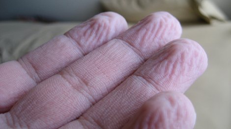
When immersed in water for a long time, the skin on your hands and feet wrinkles.
(Photo by Brenderous, click for full credit)

Almost everyone has experienced it. When you have been soaking in the bathtub, swimming, or just washing dishes for a long time, the skin on your fingers (and toes) wrinkles. From a scientific point of view, there are at least two questions to consider: (1) How does this happen? and (2) Why does this happen? Most textbooks explain (often incorrectly) the how, but I haven’t found any that explain the why. It seems that over the years, scientists have been studying this, and in the end, they have mostly answered both questions.

Let’s start with the “how.” A lot of textbooks and websites incorrectly explain this part. They say that it is the result of your skin absorbing water and swelling. It turns out that’s not true at all. More than 70 years ago, scientists showed that if certain nerves to the hand are damaged, its skin will not wrinkle, no matter how long it stays underwater.1 Over the years, other scientists have investigated water-induced wrinkling, and it seems to be the result of a process initiated by the nervous system.

Your skin is made of two layers: the epidermis (the layer you see) and the dermis (the layer underneath that contains blood vessels). When your hands and/or feet have been underwater for a long time, your nervous system tells the blood vessels in your dermis to constrict. This reduces the volume of the dermis, which in turn reduces the tension with which the epidermis is stretched. As a result, the epidermis “relaxes,” forming wrinkles.2

This answer is interesting enough, because I have long taught the incorrect explanation for how your skin wrinkles underwater. I am glad that I learned I was wrong on the point, and I will now start teaching the correct explanation. However, the “why” question is also very interesting, and a couple of recent studies have provided a good answer for that question as well.

Continue reading “Why (and How) Your Skin Wrinkles Underwater”

Human and Chimp DNA Only 70% Similar, At Least According to This Study

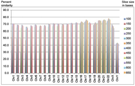
A chromosome-by-chromosome comparison of chimpanzee and human DNA. The chimp DNA was cut into slices of varying lengths (see legend on the right), and a similar sequence was searched for on the relevant human chromosome, which is shown on the horizontal axis.
(Copyright Answers in Genesis, published at http://www.answersingenesis.org/articles/arj/v6/n1/human-chimp-chromosome in a study by Jeffrey P. Tomkins)

PLEASE NOTE: The results of this study are known to be wrong due to a bug in the computer program used. A new study that uses several different computer programs shows an 88% overall similarity.

I have written about the similarity between human and chimpanzee DNA three times before (here, here, and here). It’s an important question for creationists, intelligent design advocates, and evolutionists alike, since the chimpanzee is supposed to be the closest living relative to human beings. As a result, a comparison of chimp DNA to human DNA gives us some idea of what the process of evolution would have to accomplish to turn a single apelike ancestor into two remarkably different species like chimpanzees and people.

Early on, it was widely thought that human DNA and chimp DNA were 99% similar. As I discussed in my first post on this subject, that was based on a very limited analysis of only a minute fraction of human and chimp DNA. Now that the entire set of nuclear DNA (collectively called the “genome”) of both humans and chimpanzees have been sequenced, we now know that the 99% number is just plain wrong. Interestingly enough, however, even though both genomes have been fully sequenced with a reasonable amount of accuracy, no one can agree on exactly how similar the two genomes are.

Why is that? Because comparing genomes is a lot harder than you might think. While we know the sequence of the chimp and human genomes really well, we don’t understand the DNA itself. Indeed, there are large sections of DNA that seem to be functional, but we simply have no idea what they do. As a result, comparing the genomes of two different species can be very, very tricky.

Continue reading “Human and Chimp DNA Only 70% Similar, At Least According to This Study”

Even Shark Embryos Detect Electric Fields

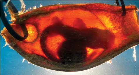
A bamboo shark embryo in its egg case (Image from the study being discussed)

I have always been fascinated by sharks. In fact, of all the times I have been scuba diving, the dive I remember the most occurred off the coast of South Africa. I went with some marine biologists who had been studying sand tiger sharks (Carcharias taurus). As I initially sunk down into the water, I leveled out just a few feet from the bottom, and as I was checking my gauge to determine my depth, a 2-meter (6-feet) long shark swum right underneath me! We ended up seeing more than a dozen sharks of various sizes on that dive. It was incredible.

One of the things that is fascinating about sharks is the way they hunt. While they use their sense of smell (and to some degree their sense of sight), one of their main hunting techniques involves using their electrical sense. Yes, sharks can sense electrical fields, and they are quite good at it. As a recently published book on sharks says:1

They can detect the minute electrical currents generated by the nervous systems of prey by using electrical sensors called the ampullae of Lorenzini…These sophisticated sensors are very useful in finding prey buried under the sand.

Interestingly enough, these sophisticated sensors often develop while the shark is still an embryo. Is that just to get the shark ready for hunting its prey when it is fully developed, or could there be some use that the embryo has for sensing electrical fields? It doesn’t need to hunt, so for what purpose could it be using its electrical sensors?

Some Australian scientists decided to investigate this issue, and what they found is fascinating!

Continue reading “Even Shark Embryos Detect Electric Fields”

Sockeye Salmon Use Geomagnetic Details to Guide Them Home

A sockeye salmon jumps a beaver dam to get back to the stream from which it hatched. (Click for credit)
Salmon have an incredible lifecycle. They hatch in freshwater streams, eat and mature a bit, and then they make their way into the ocean. They do most of their growing and maturing in the ocean, and when they are ready to reproduce, they return to the very stream from which they hatched to find a mate and begin the cycle all over again.

A lot of research has been done trying to figure out how the salmon know the way back to the specific stream from which they hatched. Well, each stream has it own mix of soil, rocks, and other environmental elements. As a result, each stream has its own specific mix of chemicals. Most of the data indicate that a salmon remembers the specific chemical makeup of its original stream. Once it gets back to freshwater, then, it starts following a “chemical trail” that will lead it back to the stream from which it hatched.1

While this is rather impressive, it doesn’t really tell us about the most difficult navigational aspect of the journey. The salmon spends a long time in the ocean growing and maturing. As a result, it often ranges far from the place where it entered the ocean. How does it find its way back to the correct freshwater inlet that will lead to the correct stream? It seems hard to believe that it could follow a “chemical trail” that far! Since we know that salmon are equipped with biological machinery that allows them to sense the earth’s magnetic field, it has been thought for some time that salmon use magnetic guidance to get them to the proper freshwater inlet.2

While that’s a reasonable conclusion based on what we know, there hasn’t been a lot of evidence to back it up. However, a recent study published in Current Biology has begun to remedy that situation.

Continue reading “Sockeye Salmon Use Geomagnetic Details to Guide Them Home”

Even More Impressive DNA Storage

DNA can store incredible amounts of information (montage of public domain images)
A few months ago, I wrote an article about a group of scientists who stored a book that contained words, illustrations, and Java script on DNA. It was an amazing technical achievement, and it demonstrated the incredible storage capabilities of this marvelous biomolecule. Well, another team of scientists has gone even further: they stored words, pictures, and audio on DNA!

Yes, the team encoded all 154 of Shakespeare’s sonnets, a photograph of the European Bioinformatics Institute (where the scientists work), and a 26-second audio clip from Martin Luther King’s famous “I have a dream” speech.1 Also, in a very fitting symbolic gesture, they added the famous James Watson and Francis Crick paper that first revealed the structure of DNA.2

This new achievement was noteworthy for more than just the fact that the scientists stored audio on DNA. While the method that the previous team used to store the book worked well, it was difficult for instruments to retrieve the information from the DNA once it was stored there. Thus, the time it took to retrieve the book from DNA storage was fairly long. The scientists who produced this study used a different method to store the information, which made it much easier for instruments to read it back. As a result, not only was everything retrieved from DNA storage with 100% accuracy, the time it took to get it back was significantly reduced.

Continue reading “Even More Impressive DNA Storage”

Being Degenerate Can Be Very Good!

DNA uses four nucleotide bases taken three at a time to code for an animo acid (click for credit).
The genetic code is degenerate, but that doesn’t mean it is immoral or corrupt. In fact, in the case of the genetic code, degeneracy is a good thing! Let me explain. One of DNA’s jobs is to tell the cell what proteins to make and how to make them. As a result, it stores “recipes” for proteins, and we call those recipes genes. Well, a protein is produced when smaller chemicals, called amino acids, are linked together in long chains that then fold into intricate shapes. So in order to tell a cell how to make a protein, a gene needs to list a string of amino acids. If the cell puts those amino acids together in the order specified by the gene, the correct protein can then be produced.

How does a gene list the amino acids? As shown in the illustration above, it does so by using the four nucleotide bases known as cytosine (C), guanine (G), thymine (T), and adenine (A). A group of three nucleotide bases codes for a specific amino acid. For example, when a gene has three thymines in a row (TTT), this means “use the amino acid called lysine.” When it has three guanines in a row (GGG), it means “use the amino acid called proline.” So by grouping its four nucleotide bases three at a time, a gene specifies which amino acid should be used in building a protein.

Here’s the catch: There are only 20 amino acids in the standard proteins of life. As a result, there need to be only 20 codes to specify them. However, there are 64 possible ways you can group four nucleotide bases three at a time. Thus, there are 64 different possibilities for how a gene can specify an amino acid, but there are only 20 amino acids the gene needs to specify. As a result, most amino acids are specified by more than one set of three nucleotide bases. As I said above, a sequence of three thymines (TTT) means “use the amino acid called lysine.” However, two thymines followed by a cytosine (TTC) means the same thing. This is why we say the genetic code is degenerate. It has multiple ways it can specify most amino acids.

Continue reading “Being Degenerate Can Be Very Good!”

Poop Transplants Treat Clostridium difficile Infections!

Clostridium difficile from a stool sample, magnified 3,006x (public domain image fron the CDC)

Clostridium difficile is a bacterium that produces toxins which can kill intestinal cells and cause severe inflammation of the intestinal walls. Mild infections from this bacterium can cause diarrhea, while severe infections can cause death. Over time, C. difficile infections have gotten worse. As one medical resource states:1

C. difficile infection has transformed from a nuisance into a potentially life-threatening illness with an attributable mortality rate of up to 16.7%.

The typical treatment for severe cases of C. difficile infection is a round of strong antibiotics, but that doesn’t always work. A significant number of patients end up experiencing one or more recurring infections within 60 days.2 As a result, medical researchers are trying to come up with new ways to treat this infection.

In a recent study published in the New England Journal of Medicine, researchers tested an…interesting…approach. They took 43 patients who had a C. difficile infection and split them into three groups. The first received the standard antibiotic treatment (in this case, 500 milligrams of vancomycin four times per day for 14 days). The second received the standard antibiotic treatment plus a bowl lavage 4 or 5 days into the treatment. In case you aren’t familiar with the term, a bowel lavage involves flushing out the intestines. It is usually done to prepare the intestine for medical imaging. The third group was given a shortened round of antibiotics (500 milligrams of vancomycin four times per day for 4 or 5 days), a bowel lavage, and then a poop transplant.

Yes, you read that right. After the bowel lavage, the patients were given a mixture of water, salt, and the feces from a healthy donor. Now don’t worry. They didn’t have to eat or even smell the stuff. It was sent into their intestine through a sterile tube that went up the nose, down the esophagus, through the stomach, and into the start of the small intestine. While the process sounds incredibly gross, the results were amazing!

Continue reading “Poop Transplants Treat Clostridium difficile Infections!”

Astronomers are Finally Starting to Question an Absurd Assumption

The black circles in this figure represent the 73 quazars that make up the largest structure in the observed universe. The red crosses represent the 34 quazars that make up another massive structure. (Image is from reference 3.)

One of the more absurd assumptions that is routinely made in astronomy is called the cosmological principle. One way to phrase the principle is:

Viewed on a large enough scale, the properties of the universe are the same no matter where you are.

However, observations have never supported this assumption. Instead, the observable universe seems incredibly “lumpy,” with huge structures separated by vast areas devoid of structures. Nevertheless, cosmologists have doggedly taken the cosmological principle as their starting assumption when it comes to developing models of the universe, despite the fact that observations don’t support it.

Indeed, the cosmological principle is a necessary starting point for the Big Bang, which most, but certainly not all, astronomers think is a good description of the origin and development of the universe. As Paul Fleisher says in his book, The Big Bang:1

The cosmological principle is the central idea of the Big Bang theory. This rule says the universe is homogeneous and isotropic at very large scales.

Even if we go away from the Big Bang model, the vast majority of models that attempt to describe the universe start with the assumption that the cosmological principle is valid. There are some models that do not start with that assumption, but they are few and far between.2

I have always been skeptical of the cosmological principle, simply because it isn’t supported by observation. The universe doesn’t look homogeneous at all. Instead, it looks really “lumpy.” Nevertheless, when I read the scientific literature, the cosmological principle seems to be considered a fact in almost all of the astronomy-related papers.

It looks like that might be starting to change.

Continue reading “Astronomers are Finally Starting to Question an Absurd Assumption”

Horizontal Gene Transfer: Another “Way Out” for Evolutionists

Horizontal Gene Transfer (represented by the arrows and paths connecting the different lineages in the drawing) is a convenient way for evolutionists to explain around the fact that the data falsify their predictions. (Click for credit)

When I was a young, impressionable student at university, I was taught as fact that all organisms on this planet could be arranged in a hypothetical “tree of life” that showed how all of them evolved from a single, common ancestor. It didn’t matter that no such tree had actually been constructed. I was told that in time, we would be able to sequence DNA quickly and efficiently, and once that happened, the tree of life would emerge from the data in all its glory. However, once DNA analysis did become reasonably quick and efficient, the tree of life never emerged. Instead, the supposed evolutionary relationships that had been determined from the fossil record were contradicted by those that were determined from the genes. Worse yet, the genetic story of evolution changed depending on which specific genes were studied.

This was especially apparent in the analysis of single-celled organisms. As more and more genetic analyses were done on such organisms, it became increasingly obvious that there was simply no way to arrange their genetic information into any pattern that even remotely resembled a hypothetical tree of life. Some scientists understood what this meant: there is something seriously wrong with the evolutionary framework to which biologists have been clinging. As a result, they have started investigating other, more promising paradigms such as creationism or intelligent design. However, most biologists continue to cling to a view that has been falsified again and again by various data. As a result, they had to find a “way out.” They had to find some means by which they could explain around the fact that the genetic data falsified the tree of life.

Enter the concept of Horizontal Gene Transfer, also known as lateral gene transfer. In this process, a section of DNA is transferred from the genome of one organism to the genome of another, unrelated organism. In other words, rather than passing down a gene through the process of reproduction, horizontal gene transfer allows a gene to travel horizontally between unrelated organisms.

Now the phenomenon itself was known long before the problem with the tree of life had been documented. Way back in 1960, for example, Japanese scientists determined that an antibiotic-resistant bacterium could transfer its resistance to an unrelated bacterium that was not resistant to the antibiotic.1 In time, the mechanism was fully worked out, and it was demonstrated that bacteria could, indeed, swap genes back and forth. As it became increasingly clear that this was a common phenomenon among bacteria, it was recognized that horizontal gene transfer could “smear out” the tree of life for single-celled organisms.2

Since horizontal gene transfer was so successful at explaining away the failed evolutionary prediction of the tree of life when it came to single-celled organisms, it’s not surprising that this same “way out” is now being used to explain why certain genes in animals do not fit the pattern predicted by the evolutionary hypothesis.

Continue reading “Horizontal Gene Transfer: Another “Way Out” for Evolutionists”

Male DNA in Female Brains? Yes!

Male DNA was found in 63% of the women studied.
(Public domain image)
A gene called DYS14 is found only on the Y chromosome in human beings. Of course, only males have a Y chromosome, so it is reasonable to assume that the only place you will ever find this gene is in men, right? Wrong! William F. N. Chan and his colleagues examined the brains of 59 deceased women, and they found the gene residing in 37 of the brains studied! In other words, 63% of the deceased women studied had male DNA in their brains. Interestingly enough, in most of those brains, the DNA was found in several different places!1

How in the world did male DNA get into these women’s brains? The researchers aren’t sure, because they don’t have detailed medical histories for most of the women. However, the most likely explanation is that the DNA comes from the male children that these women carried. I have written about this phenomenon, called fetomaternal microchimerism, before. As I mentioned in that article, when I first heard about a baby leaving cellular remains inside his or her mother, I thought it couldn’t possibly be true. However, I was wrong. There is solid evidence to suggest that not only do babies leave a lasting, cellular imprint on their mothers, mothers do the same for their babies.

However, the possibility that children leave some of their DNA behind in their mother’s brain is very surprising. After all, the cells that make up the brain are incredibly sensitive. In fact, the contents of your own blood are toxic to your brain cells. As a result, you have an elaborately designed blood-brain barrier that shields your brain cells from your blood. This barrier is so vigilant that it allows only certain substances (such as the glucose and electrolytes that the brain cells need) to pass through it. As a result, your brain cells are protected from the majority of substances found in your bloodstream.2

Continue reading “Male DNA in Female Brains? Yes!”