A Bacterium That “Eats” Carbon Dioxide…and a Creationist Prediction

A false-color scanning electron microscope image of Escherichia coli. The different colors represent bacteria with different traits.

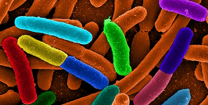
Escherichia coli (E. coli) are the workhorses of the bacterial world. They are found in every human being and most warm-blooded animals, but they are also found in laboratories all over the world. Because they are easy to care for, reproduce quickly, and have a genome that is reasonably well-understood, they are a popular subject of study among biologists. In addition, they end up producing a lot of chemicals that we need but are unable to produce ourselves. For example, insulin is a protein that all people need, but some people don’t make enough of it (or don’t respond well enough to it) to remain healthy. That leads to diabetes, and one treatment for diabetes is regular insulin injections.

While the insulin in pigs and cattle is close to what we find in people (and was used to treat diabetes for a long time), the best insulin for most diabetics is human insulin. Unfortunately, even with the best technology available, we aren’t good enough chemists to make insulin, but simple organisms like bacteria are. As a result, scientists have learned how to insert the human gene for insulin into a bacterium, which allows the bacterium to do the chemistry for us. As a result, much of the insulin used to treat diabetics today is human insulin produced by E. coli bacteria.

Unlike some species of bacteria, however, E. coli have to eat in order to get the energy and raw materials they need to do that chemistry. This ends up producing carbon dioxide as waste. To reduce the buildup of carbon dioxide in the atmosphere, then, it would be nice to produce chemicals like insulin from an organism that does not produce carbon dioxide in order to live. There are technical problems with that, however, so right now, diabetic insulin (and many other medically-related chemicals) adds to humanity’s “carbon footprint.” There are two ways to fix this: Either figure out how to use organisms that don’t have to eat (like organisms that make their own food through photosynthesis) or change E. coli so that it doesn’t have to eat.

In a recent study, scientists have been working on the second alternative and have experienced some success. As a byproduct, they have produced something that can be used to test creationism.

Continue reading “A Bacterium That “Eats” Carbon Dioxide…and a Creationist Prediction”

When Is a Person Actually Dead?

A student recently sent me an article from Live Science that reports on a man who was declared dead by three doctors. Four hours later, as he was being prepped for an autopsy (the marks to guide the autopsy had already been put on him), he started snoring! As of the time the article was written, he was alive and in the intensive care unit of a hospital. The student asked how such a thing could happen. Was it incompetence on the part of the doctors, or is it difficult to tell whether or not a person is dead? I told the student that while I couldn’t address the details of this particular case since I wasn’t involved, I could tell him that there have been cases over the years where the experts were convinced that a person was dead when, in fact, that person wasn’t.

I first heard this kind of story when I was preparing for a talk about miracles. I ran across the case of Emma Brady. She had been declared dead after exhibiting no vital signs. She was placed in a body bag and taken to the morgue. When her children arrived about an hour later to say their goodbyes, they found her gasping for air. The administrator of the hospital said that after the family told a nurse about what they had seen:

Miraculously, the patient exhibited vital signs that were absent previously.

Over the years, I have kept my eye out for stories like this, and while they are rare, they are most certainly not unheard of.

Consider, for example, the story of Steven Thorpe. At age 17, he was in a tragic accident that killed one of the other occupants of the automobile. He was put in a medically-induced coma, and a team of four physicians told his parents that he was brain dead. They suggested that his organs be donated to help others. However, the parents brought in an additional doctor (a neurologist), who demonstrated faint brain activity. The doctors at the hospital agreed to bring him out of the medically-induced coma, and Thorpe recovered. He left the hospital five days later and at the time the article was published, he was alive and well.

Once again, while these stories seem rare, they are not unheard of. In 2008, Zack Dunlap was in an automobile accident and was declared dead 36 hours later. However, he wasn’t dead. In fact, he says that he actually heard his doctors saying that he was dead. The hospital made plans to harvest his organs, since his driver’s license said that he was an organ donor. However, as his family was saying goodbye, one of his cousins (a nurse) decided to pull out his pocket knife, hold Zack’s foot, and scrape the knife against it. Zack pulled his own foot out of his cousin’s hand. The family took it as a sign of life, and they argued that the hospital should treat Zack as if he were alive. He ended up making a full recovery.

The bottom line is that while we have amazing technology and a lot of knowledge about human anatomy and physiology, there are limits to what we can detect and what we can conclude. While it is sometimes very obvious that someone is dead, there are other times when even the experts can be fooled. That’s something all of us need to keep in mind when we deal with life and death issues.

No, It’s Not a Picture of an Atom

When I wrote the first edition of my high school chemistry textbook (back in 1994), I discussed the impossibility of seeing atoms. Atoms aren’t just small; they are simply too small to be seen, even with the most powerful microscope. That’s because in order for us to see something, light must bounce off it and enter our eyes. Cells on our retina detect that light, send nerve signals to our brain, and our brain interprets the signals to form an image. Since atoms are roughly 1,000 times smaller than the smallest wavelength of visible light, there is no way for light to bounce off a single atom. As a result, we will never be able to see individual atoms, no matter how powerful a microscope we use.

I said that every year to every general chemistry class I taught at the university level, and no one ever questioned my assertion. But homeschoolers think more critically than most students, so the very first year my chemistry course was published, I got a FAX (yes, a FAX) from a student questioning my statement. The student showed me an image like the one below, which was taken with a scanning tunneling electron microscope at IBM. She asked if it wasn’t a picture of individual atoms:

An image of the surface of nickel, as produced by a scanning tunneling electron microscope (click for source)

While the image is generally presented as a “picture” of nickel atoms, it is not. It is a graph that represents data that have been processed through a series of equations which govern the behavior of electrons traveling at high speeds. The equations depend on multiple theories, including special relativity and quantum mechanics. I am pretty confident that those theories are mostly true, so I am pretty confident that this is a realistic representation of atoms on the surface of a nickel foil. However, it is not a picture. Light was not used (high-energy electrons were used), and the image is not a direct representation of the nickel surface. It is a direct representation of data that have been processed through mathematical equations that come from several theories.

Recently, I got a question from a teacher who is using my new chemistry course. A student brought him an article that says scientists have been able to take a picture of a single strontium atom. I told the teacher why it is not a picture of a strontium atom, and then over the weekend, I got the same question on my Facebook page. As a result, I decided to write a quick article (well, as quick an article as I can write) about it.

This is definitely a picture, as it is the result of directly detecting light. And, that light is coming from a single atom. That’s the remarkable aspect of the photograph. Because atoms are so small, it is very hard to isolate a single one, but the experimenter who took the picture was able to do just that. However, it is not a picture of the atom. It is a picture of the visible light that the atom is emitting as a result of being excited. When atoms are excited, they must get rid of that excess energy, and one way they can do that is to emit light. The strontium atom is emitting visible light, which is why a picture of it could be taken.

Once again, however, this is not a picture of the atom; it is a picture of visible light being emitted by the atom. After all, the picture shows that the light is purple. If this were a picture of a strontium atom, it would mean that strontium atoms are purple. However, atoms have no color, because they are smaller than the smallest wavelength of visible light.

If you think I am just being pedantic here, consider the picture below:

Tubes of neon atoms emitting light as a result of being excited by electricity. (click for credit)

Each tube in the picture contains neon gas, and electricity is being passed through the gas, giving the atoms extra energy. To release that energy, they emit light, and the visible wavelengths that are emitted produce the orange color that you see in the picture. Are you seeing all the individual neon atoms in those tubes? Of course not. You are seeing the light that those excited neon atoms are emitting.

The “picture” of the strontium atom is the same thing, except that there is only one atom emitting light, while there are roughly 100,000,000,000,000,000,000,000 neon atoms emitting light in the picture above.

Incredibly Fragile Dinosaur Soft Tissue

Two images of the delicate, one-way valves from veins. They were found in dinosaur soft tissue!
(Image copied from the presentation embedded below)

Mark Armitage and James Solliday at the Dinosaur Soft Tissue Research Institute have been doing some amazing work. On October 5th, Mr. Armitage presented their findings at Lower Columbia College. Apparently, he has not yet received the video of that presentation, so he kindly posted a quick overview of the content. To me, it is astounding:

While everyone should watch all 15 minutes of the presentation, I want to highlight the things that I think are most important.

At 2:29, he shows two images that elicited an audible gasp from me when I first saw them. To understand just how incredible the images are, you need to know that there are one-way valves found in vertebrate veins. This is because the blood pressure in a vein is so low that blood can actually travel backwards. To prevent that, there are delicate, one-way valves throughout the veins. They open when the blood is flowing the correct way, and they close to prevent it flowing backwards. In the left-hand part of the image at the top of the post (copied from the presentation), you see a circle with what looks like a partially-opened tent flap. The circle is the base of the valve, and the “tent flap” is the delicate membrane that opens and closes. In that image, the valve is partly open. On the right-hand side, the valve is fully open.

This is incredible to me, because I have tried to dissect animals and extract these valves. I have never been able to. They are so delicate that I end up destroying them in the dissection process. Now, of course, I am not much of a biologist, and I am even less of an expert at dissection. Nevertheless, my experience with them indicates that they are absurdly delicate. Yet, here they are in a dinosaur fossil! Not only does this give evidence that the fossil is not millions of years old, but it also shows that these are definitely not structures that come from fungi or bacteria which recently invaded the fossil. Bacteria and fungi do not build structures with these delicate, one-way valves! He also presents other evidence that rules out bacterial and fungal contamination.

At 8:22, he shows red blood cells from a fossil that is supposed to be 400 million years old! The cells have the appropriate size and shape for red blood cells. Later on (12:05), he shows a blood vessel from a dinosaur fossil that has not even collapsed! It has an air bubble in it. When he does a stain test to see what is in the blood vessel, the test indicates that there is RNA in the blood vessel!

At 6:47, he shows what appears to be blood clotted in the tissue. He shows how it behaves just like you would expect blood to behave when exposed to polarized light, and he also shows that iron from the blood has not spread into the bone tissue. This is important, because Dr. Mary Schweitzer has proposed that iron might be preserving the soft tissue found in dinosaur bones. There has already been several arguments (see here and here) that seem to invalidate Dr. Schweitzer’s hypothesis, but this observation is the nail in the coffin. Iron can’t be preserving bone tissue if it doesn’t spread into the bone to begin with!

I have said this before and will say it again: It’s a wonderful time to be a young-earth creationist!

NOTE: A commentor made the great suggestion that I post a link if you want to support Mr. Armitage’s research. Here it is:

Donate to the Dinosaur Soft Tissue Research Institute.

Rat Surgeons?

I have written previously about Australia’s cane toad problem (here and here). In 1935, cane toads were brought in to control a pest that was feeding on sugar cane in northeastern Queensland. They ended up being ineffective at controlling the pest, and because they have few natural predators there, Australia was ineffective at controlling them. They have been spreading out across Australia since 1935, and there is no end in sight to their population’s expansion.

As I have discussed previously, cane toads have already affected wildlife in the areas where they have become established. Because they are large (for toads) and the adults are poisonous to snakes, for example, the average head size of a snake has decreased in those areas with a significant cane toad population. After all, the snakes that have large enough heads to eat the adult toads die. As a result, snakes that can’t eat them (snakes with smaller heads) are significantly more likely to survive. They survive because they cannot eat what would kill them!

But there is another way to survive in the presence of cane toads: figure out a way to eat them without being poisoned. Based on the results of a recent study, it seems that one clever Australian predator has learned to do just that. The authors of the study were intrigued when they started finding cane toad bodies that had what appeared to be surgically-precise incisions on their bodies. They eventually set up some infrared cameras and found that golden-bellied water rats were the ones making the incisions.

It turns out that only the skin and certain organs (like the bile duct) in the frog are poisonous. If a predator can avoid those structures, it can eat the toads without being harmed, and apparently, the water rats have figured that out. The researchers found that the heart and liver had been removed in each dead cane toad, presumably eaten by the rats. In the largest toads, the skin of the legs was also peeled back and the leg muscles were eaten. The authors say that all of this was done with a high level of precision.

The question, of course, is how the rats figured this out. The researchers are not sure. They know that water rats feed on other toad species as well as the younger cane toads that aren’t as poisonous, and it may be that in this area, that’s the way rats eat all the toads they kill. It’s also possible that some rats just stumbled onto this technique and passed it on to their offspring. As the authors note, water rats care for their offspring for weeks after they have been weaned, so it would be easy for the young rats to learn how their parents are eating the toads. The researchers note that for now, this feeding technique is limited to the water rats in certain areas, but they suggest that it might spread as time goes on.

Add the Australian water rat to the ever-growing list of surprisingly clever animals (see here, here, and here.)

Why Do I Include God in My Science Texts?

13th-century illumination from the French Bible Moralisée, depicting Christ (who is God) creating the World.
I was asked that question a couple of days ago, but it wasn’t the first time. Over the years, several people have asked me why I write about God in my science textbooks. After all, science is about facts, while belief in God is about faith. Science doesn’t belong in religious texts, and God doesn’t belong in science texts. That may sound reasonable in today’s world, but it is simply dead wrong. In addition, it demonstrates a shocking level of ignorance about the history of science. Nevertheless, it is a good question, and it deserves a detailed answer.

First, there is an obvious philosophical reason: God is the source of all that science studies, so it only makes sense to discuss Him in the context of studying His creation. Consider, for example, teaching a course on U.S. Law. Would you try to teach it without referring to the U.S. Constitution? Perhaps. Maybe there are law professors who do just that, but for me, I can’t imagine discussing U.S. law without discussing the source from which it comes. In the same way, I find it pointless to discuss science without discussing its source.

Second, there are practical reasons to discuss God while teaching students about science. If we emphasize the fact that the things we study as scientists are designed, we give students a superior way in which to view the natural world. Those who want to reject the idea of a Creator God will try to convince students that this world was “thrown together” by random chance. As a result, students get the idea that creation is full of shabby constructions. Of course, nothing could be further from the truth. The designs found in the natural world make our best technology look like garbage. This has led one desperate atheist to write:1

Biology is the study of complicated things that give the appearance of having been designed for a purpose.

Of course, a more reasonable evaluation of the data leads us to the conclusion that biology is the study of things that have been designed for a purpose.

This is very important, because when we understand that biology is the study of designed things, we don’t fall prey to misconceptions that hold back the progress of science. How many lives have been wasted because scientists looked at the primary cilium as a evolutionary vestige rather than an antenna designed to receive signals? How long did scientists delay a more detailed understanding of genetics because of the nonsensical notion of “junk DNA”? How many people needlessly suffer relapses of intestinal bacterial infections because of the silly idea that the appendix is a vestigial organ? A scientist who understands that the natural world is designed is simply better able to interpret what he or she is studying, since it is the more realistic view.

Finally, there is a spiritual reason to include God in science. As Nobel laureate Dr. Arthur Leonard Schawlow once put it:2

But the context of religion is a great background for doing science. In the words of Psalm 19, “The heavens declare the glory of God and the firmament showeth his handiwork.” Thus scientific research is a worshipful act, in that it reveals more of the wonders of God’s creation.

Ever since I became a Christian, science has been a worshipful act for me. There is no way I could write about this worshipful act without including the One who is being worshipped!

REFERENCES

1. Dawkins, R., The Blind Watchmaker, W.W. Norton & Company, New York, USA, p. 1, 1986.
Return to Text

2. Cosmos, Bios, Theos, Henry Margenau and Roy Abraham Varghese, ed., Open Court Publishing 1992, p. 106.
Return to Text

What’s Really Causing Coral Bleaching?

The soft coral colony at the center of the picture is bleached. The corals to the right are not. (click for credit)

Coral are amazing animals. They live in a mutualistic relationship with algae, giving the algae a safe home in exchange for some of the food that the algae make through photosynthesis. The variety of colors seen in a coral reef are a result of this relationship. However, coral sometimes expel their algae, turning white. This is called “coral bleaching,” and it generally happens when the water is warmer than usual. the Australian Marine Conservation informs us:

Coral bleaching is a global crisis, caused by increased ocean temperatures driven by carbon pollution.

This has become a common mantra in the “global warming is going to kill us all” movement, because coral reefs are so fundamentally important to the health of ocean ecosystems. Indeed, it has become so important that if you question what the global-warming alarmists say, it can lead to dire consequences.

Consider, for example, the case of Dr. Peter Ridd. Some of his colleagues at James Cook University published work indicating that Australia’s Great Barrier Reef was on the verge of collapse because of global warming. Dr. Ridd dared to question that narrative, pointing out the data that indicate there is nothing unusual about the bleaching events that have been occurring at the Great Barrier Reef and that the reef has about the same amount of healthy coral as it did back in 1985. For that transgression, he was fired. While a court has ruled the firing unlawful, the university plans to appeal the ruling. Regardless of what happens at appeal, it is clear that the firing was anti-science. Criticism of data, even data related to sacred cows such as global warming, is the hallmark of good science. To squelch such criticism is a direct assault on the progress of science.

Of course, the real question is whether or not global warming is a threat to the oceans’ coral reefs. The answer remains unclear, but the balance of the evidence indicates that it is not. For example, one study of the Great Barrier Reef shows that bleaching events were more common several hundred years ago. According to that study, bleaching events hit their peak in the 1850s. There is also some indication that bleaching is an adaptive mechanism and is not necessarily bad for the health of a coral reef.

Continue reading “What’s Really Causing Coral Bleaching?”

Something I (and Most Scientists) Have Taught for Many Years is Probably Wrong!

One of the problems that science textbook authors face is the fact that science is constantly changing. As we learn more about the Creator’s handiwork, we find that the science we have taught as fact is actually incorrect. Sometimes, this is because the experiments upon which those facts are based were in error. Sometimes, it’s because our interpretations of those experiments were in error. Sometimes, it’s a result of making conclusions that go beyond what the experiments actually tell us. The practical upshot of all this is that some of the things you are reading in your science textbooks are wrong.

I recently found out that something I (and most other authors) have taught about DNA is probably wrong. Most people know that DNA is a double helix. As shown in the illustration above, those two helixes wind around each other, with the information-bearing units (called nucleotide bases) inside. In order for cells to use the information in DNA, those helixes have to be separated so that the sequence of the nucleotide bases can be read. That means the helixes need to be held together when DNA is not being used, and then they must be separated when it is time for the cell to read the DNA.

How does that happen? Well, according to most textbooks (including mine), it is because the nucleotide bases form hydrogen bonds with one another. Hydrogen bonds are weaker than true chemical bonds, but they can hold things together. As I say it in my textbook, Exploring Creation with Biology, 2nd Edition:

…the attraction between the atoms in hydrogen bonding is about 15% as strong as the attraction between two atoms that have a true chemical bond linking them. Thus, the hydrogen bonds in DNA are strong enough to keep the two chains together in a double helix, but they are significantly weaker than a true chemical bond. Since they are weaker than a true chemical bond, it is rather easy for the two helixes in DNA to unravel.

This sounds great, but a recent study indicates that it’s probably not true. If nothing else, it doesn’t tell the entire story.

Continue reading “Something I (and Most Scientists) Have Taught for Many Years is Probably Wrong!”

Large Study Indicates Genetics Has Little Influence on Sexual Orientation

I don’t normally write on topics like this, because studying human behavior is a tricky subject. There are all sorts of different explanations for a given behavioral characteristic in people, and trying to isolate a specific cause is difficult, to say the least. However, there has been a lot of news about the recent study that concluded there is “no gay gene,” and I have gotten several questions about it. As a result, I decided to read the study and share my thoughts.

First of all, it’s not surprising that there is no gay gene. In fact, researchers have said that for years. If there were a single gene that heavily influenced whether or not a person is homosexual, it would have been easy to find and discovered years ago. Also, even something as simple as the color of your eyes is governed by at least eight different genes. Thus, to think that something as complex as sexual behavior is governed by one gene is naive at best. So that specific result of the study is not even interesting, much less newsworthy. What makes the study newsworthy is its size, its scope, and the fact that its conclusions are very weak.

The study is massive in two ways. First, the main study looked at 477,522 individuals, but then it repeated the study using three smaller datasets that were composed of 15,142 individuals. Whenever you study people, the more people you have, the less uncertain your results will be. Thus, the sheer number of individuals in the study makes it important. Second, the study compared the entire genomes of the individuals. In other words, they looked at all the DNA found in the nucleus of the individuals’ cells. That’s a massive amount of data for a massive number of people!

What it tried to do is compare single nucleotide polymorphisms (SNPs) among the individuals to see if they could be correlated with sexual behavior. If you aren’t familiar with the term, SNPs are the most common variation between sets of human DNA. Genetic information comes in sequences of DNA building blocks called nucleotide bases. There are roughly three billion nucleotide bases in one strand of human DNA. An SNP is a change in one of those nucleotide bases.

Continue reading “Large Study Indicates Genetics Has Little Influence on Sexual Orientation”

Excessive Screen Time Linked to Poor Psychological Health in Children and Adolescents

A child playing with an Apple iPad
(click for credit)
Screens are everywhere. It’s very hard to avoid them. However, many health organizations recommend that parents limit the amount of screen time children and adolescents have. First, the health effects of excessive screen time have been well documented. Children who watch TV, computer, phone, and tablet screens a lot don’t exercise much. That leads to some very poor health outcomes, such as obesity. Second, a recent study indicates that there are significant negative psychological effects associated with a lot of screen time.

Psychological research is difficult for many reasons. Whenever you deal with people, you have to try to control for all sorts of variables that affect each subject, and those variables are often significantly different in different subpopulations. In addition, some of outcome measures are subjective, at best. As a result, psychological studies often have conflicting results. However, this one seems to do a good job getting over those hurdles. It contains a lot of subjects (40,337) who were randomly selected, leading to an averaging-out of at least some of the variables. Also, it used the results of the National Survey of Children’s Health (NSCH), which was done in 2016. This survey was given to the people who know the subjects best: their caregivers. It also asks a lot of fairly objective mental health questions, such as whether or not the subject was ever diagnosed with anxiety or depression.

The survey also asked the subjects’ caregivers the following questions:

a) On an average weekday, about how much time does [child’s name] spend in front of a TV watching TV programs, videos, or playing video games?

b) On an average weekday, about how much time does [child’s name] spend with computers, cell phones, handheld video games, and other electronic devices, doing things other than schoolwork?

The researchers added the results of both questions to get the total amount of screen time the subject has each weekday. They then correlated that number to the mental-health-related questions on the survey. The results were rather alarming.

Continue reading “Excessive Screen Time Linked to Poor Psychological Health in Children and Adolescents”